. 2009 May 8;284(19):12862-73.
doi: 10.1074/jbc.M805123200. Epub 2009 Feb 24.

The serine protease plasmin cleaves the amino-terminal domain of the NR2A subunit to relieve zinc inhibition of the N-methyl-D-aspartate receptors

Affiliations

The serine protease plasmin cleaves the amino-terminal domain of the NR2A subunit to relieve zinc inhibition of the N-methyl-D-aspartate receptors

Hongjie Yuan et al. J Biol Chem. .

Abstract

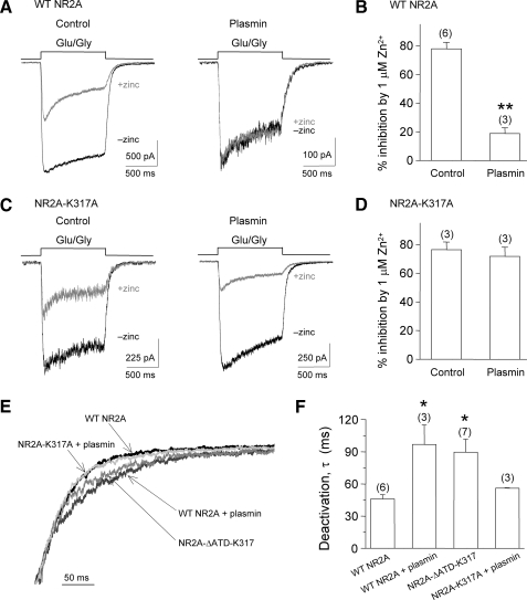
Zinc is hypothesized to be co-released with glutamate at synapses of the central nervous system. Zinc binds to NR1/NR2A N-methyl-d-aspartate (NMDA) receptors with high affinity and inhibits NMDAR function in a voltage-independent manner. The serine protease plasmin can cleave a number of substrates, including protease-activated receptors, and may play an important role in several disorders of the central nervous system, including ischemia and spinal cord injury. Here, we demonstrate that plasmin can cleave the native NR2A amino-terminal domain (NR2A(ATD)), removing the functional high affinity Zn(2+) binding site. Plasmin also cleaves recombinant NR2A(ATD) at lysine 317 (Lys(317)), thereby producing a approximately 40-kDa fragment, consistent with plasmin-induced NR2A cleavage fragments observed in rat brain membrane preparations. A homology model of the NR2A(ATD) predicts that Lys(317) is near the surface of the protein and is accessible to plasmin. Recombinant expression of NR2A with an amino-terminal deletion at Lys(317) is functional and Zn(2+) insensitive. Whole cell voltage-clamp recordings show that Zn(2+) inhibition of agonist-evoked NMDA receptor currents of NR1/NR2A-transfected HEK 293 cells and cultured cortical neurons is significantly reduced by plasmin treatment. Mutating the plasmin cleavage site Lys(317) on NR2A to alanine blocks the effect of plasmin on Zn(2+) inhibition. The relief of Zn(2+) inhibition by plasmin occurs in PAR1(-/-) cortical neurons and thus is independent of interaction with protease-activated receptors. These results suggest that plasmin can directly interact with NMDA receptors, and plasmin may increase NMDA receptor responses through disruption or removal of the amino-terminal domain and relief of Zn(2+) inhibition.


Figures

FIGURE 1.
FIGURE 1.
Plasmin cleavage of the NR2A subunit in brain tissue. A, Western immunoblot was probed with a COOH-terminal NR2A antibody (Upstate Biotech). Rat brain membranes were incubated with plasmin for 20 min at room temperature prior to loading in a 10% Tris glycine gel. Note that plasmin cleaves a ∼40-kDa fragment from the NR2A amino-terminal domain. B, arrow indicates approximate plasmin cleavage site on NR2A subunit. Line above the COOH-terminal side indicates antibody epitope.
FIGURE 2.
FIGURE 2.
Determination of the cleavage site of the NR2A subunit. A, schematic diagram of NR2A. Upper panel, histidines in the amino-terminal domain are indicated as gray ovals. The four putative membrane-associated domains are indicated as M1–M4. His42, His44, and His128 are critical for high-affinity voltage-independent Zn2+ inhibition of NR1/NR2A receptors and are highlighted by asterisks (11). The NR2AATD construct (residue Ala33–Asp421; dot boxed) was cloned into pGEX-KG vector. Middle panel, the expressed GST-NR2AATD fusion protein was COOH-terminal tagged with c-Myc (∼74.3 kDa). The vector-encoded thrombin cleavage site is indicated by an arrow. Lower panel, thrombin-cleaved NR2AATD (46.3 kDa). B, Coomassie-stained PVDF membrane containing NR2AATD protein fragments. Arrows indicate the potential plasmin (10 nm)-cleaved fragments subjected to amino-terminal sequencing. Only the band indicated by the thick red arrow yielded an interpretable sequence. C, amino acid Lys317 in red indicates the plasmin cleavage site. Note that the underlined amino acids were determined from amino-terminal peptide sequencing on the cleaved protein fragment (the bottom protein fragment in panel B, indicated by the red arrow). D, diagram of the NR2A subunit shows autonomous glutamate and Zn2+ binding domains. E, homology model of the NR2AATD. Left and middle panels, plasmin cleaves (indicated by the red arrow) between Lys317 (CPK colors) and Ala318 (first residue loop of red portion) in the NR2AATD protein (initiating methionine is 1). Zn2+ and its coordinating residues are indicated in the left panel (arrow). In the right panel, the red amino acids are downstream of the plasmin cleavage site and presumably remain attached to the receptor. The light gray portion of the protein harboring the Zn2+ site is free to dissociate from receptor after plasmin treatment.
FIGURE 3.
FIGURE 3.
A, recombinant NR2AATD proteins bind to Zn2+-charged IDA-agarose (lanes FT1 and FT2 under + zinc), and can be eluted in the flow-through in uncharged IDA-agarose (lanes FT1 and FT2 under - zinc). When the NR2AATD-bound Zn2+-IDA-agarose was eluted with 50 mm EDTA, all bound NR2AATD protein appeared in the elutes (lanes E1 and E2 under + zinc). NR2AATD proteins were probed with an anti-c-Myc antibody (1:10,000, Sigma) against the COOH-terminal c-myc inserted into the NR2AATD construct. All experiments (n = 2) were performed in duplicate (e.g. FT1 and FT2, E1 and E2). The right panel shows a shorter exposure of the same Western blot in the left panel to highlight that NR2AATD proteins that were eluted as a clear single band. M, protein standards (kDa). W1, first wash after incubation of NR2AATD and Zn2+-IDA-agarose. B, a competitive Zn2+-IDA-agarose binding assay showed NR2AATD proteins containing H44G and H128A double mutations bound weaker than the wild-type NR2AATD when challenged with 2 low affinity divalent chelating agents, imidazole and l-histidine. The immunoblot was labeled with the anti-c-Myc antibody (1:10,000, Sigma). L, WT, NR2AATD. FT, flow-through. MT, NR2AATD-(H44G/H128A). E, elute with consecutive agent. Concentrations of each agent are indicated.
FIGURE 4.
FIGURE 4.
Recombinant NR1/NR2A-ΔATD-K317 is functional and Zn2+ insensitive. A, NR2A-ΔATD-K317 was constructed by inserting an NruI site into wild-type NR2A and subcloning the downstream portion (NruI-NotI) into the vector of NR2B-ΔATD-M394. B, mean normalized concentration-response curves for glutamate (left panel) and glycine (right panel) in oocytes expressing wild-type NR1/NR2A and NR1/NR2A-ΔATD-K317. Note that the EC50 of glutamate of NR2A-ΔATD-K317 (dashed line, 3.3 ± 1.4 μm, n = 6) is similar to the EC50 of glutamate of wild-type NR2A (solid line, 5.2 ± 0.3 μm, n = 6), whereas the EC50 of glycine of NR2A-ΔATD-K317 (dashed line, 2.5 ± 0.2 μm, n = 12) is slightly shifted to the right compared with wild-type NR2A (solid line, 1.3 ± 0.1 μm, n = 14). C, two-electrode voltage-clamp recordings of oocytes injected with wild-type NR1/NR2A and NR1/NR2A-ΔATD-K317 cDNA are shown. Left, a representative two-electrode voltage-clamp current recording obtained from wild-type NR1/NR2A showed significant inhibition by 1 μm Zn2+. Right, a representative recording of currents from NR2A-ΔATD-K317 exhibited less inhibition by 1 μm Zn2+. D, mean normalized concentration-response curves for zinc inhibition in oocytes expressing wild-type NR1/NR2A and NR1/NR2A-ΔATD-K317. Note that the Zn2+ concentration-inhibition curve at wild-type NR1/NR2A receptors (n = 8) was biphasic, with high (IC50, 46.5 nm; Hill slope, 0.9) and low (IC50, 10.5 μm; Hill slope, 2.4) affinity components, whereas NR1/NR2A-ΔATD-K317 receptors (n = 8) only showed a low affinity component (IC50, 13.5 μm, Hill slope, 1.7).
FIGURE 5.
FIGURE 5.
Plasmin treatment relieves Zn2+ inhibition in whole cell voltage-clamp current recordings from HEK 293 cells expressing recombinant NR1/NR2A NMDA receptors. A, whole cell voltage-clamp recording of HEK 293 cells transfected with wild-type NR1/NR2A. Left, representative traces of currents elicited by 50μm glycine and 50μm glutamate in 10μm EDTA (black, -zinc) superimposed on currents evoked by 50μm glycine and 50 μm glutamate in 1 μm Zn2+ (gray, +zinc). Right, current evoked by 50 μm glycine and 50 μm glutamate in 10 μm EDTA (black) and 50 μm glycine and 50 μm glutamate in 1 μm Zn2+ (red) following 100 nm plasmin treatment. B, 1 μm Zn2+ inhibited HEK 293 cells expressing wild-type NR1/NR2A by 78 ± 4.6% (n = 6) but only inhibited HEK 293 cells by 19 ± 13% following a 10-min treatment with 100 nm plasmin (n = 3). **, p < 0.001; unpaired t test. C, whole cell voltage-clamp current recordings from HEK 293 cells transfected with NR1/NR2A-K317A. Left, representative traces of currents evoked by 50 μm glycine and 50 μm glutamate in 10 μm EDTA (black) and 50 μm glycine and 50 μm glutamate in 1 μm Zn2+ (gray). Right, representative traces of currents evoked by 50 μm glycine and 50 μm glutamate in 10 μm EDTA (black) and 50 μm glycine and 50 μm glutamate in 1 μm Zn2+ (gray) following 10 min treatment with 100 nm plasmin. D, the summary shows that 1 μm Zn2+ inhibited HEK 293 cells expressing NR1/NR2A-K317A by 77 ± 5.3% (n = 3) and caused 72 ± 6.5% inhibition following 10 min of 100 nm plasmin treatment (n = 3). E, representative traces of the deactivation of normalized current responses evoked by removal of glutamate from HEK 293 cells expressing NR1/NR2A (black), plasmin-treated NR1/NR2A (dark gray), NR1/NR2A-ΔATD-K317 (gray), and plasmin-treated NR1/NR2A-K317A (light gray). F, the deactivation time constants for NR1/NR2A following plasmin treatment (τ = 96.8 ± 18.1 ms; n = 3) and the deletion mutant NR1/NR2A-ΔATD-K317 (τ = 89.5 ± 12.1 ms; n = 7) were similar and statistically longer than untreated NR1/NR2A (τ = 46.2 ± 3.9 ms; n = 6). Plasmin-treated NR1/NR2A-K317A (τ = 56.0 ± 0.35 ms; n = 3) also deactivated rapidly. *, p < 0.05, one-way analysis of variance with Tukey's post-hoc test.
FIGURE 6.
FIGURE 6.
Plasmin treatment relieves the Zn2+ inhibition on NMDA receptors in cultured cortical neurons. A, the figure shows a recording pipette on a representative cultured cortical neuron. B, a representative inward Na+ current is shown in whole cell mode in response to voltage steps from -90 to +50 mV. C, extracellular 1 μm Zn2+ inhibited agonist-evoked currents in wild-type (WT) neurons (50 μm NMDA and 50 μm glycine) by 53 ± 2.6% (n = 6) in the presence of 20 μm CNQX and 3 μm ifenprodil (upper panel), but had less effect (29 ± 4.2%, n = 7) after treatment with 300 nm plasmin (lower panel). D, summary of the Zn2+ inhibition on the wild-type control and plasmin treatment group. *, p < 0.05, unpaired t test.
FIGURE 7.
FIGURE 7.
The reduction of Zn2+ inhibition on NMDA receptors of cultured cortical neurons by plasmin is independent of protease-activated receptors. A, extracellular 1 μm Zn2+ inhibited NMDA-evoked whole cell currents in PAR1-/- neurons by 40 ± 4.3% (n = 10) in the presence of CNQX and ifenprodil (upper panel), but had less effect after treatment with 300 nm plasmin (18 ± 4.0%, n = 6; lower panel; **, p < 0.001, unpaired t test, compared with the PAR1-/- control). B, summary of the Zn2+ inhibition of NMDA current responses in PAR1-/- control neurons and the plasmin treatment group of PAR1-/- neurons. C, summary of the Zn2+ inhibition of NMDA current responses on the wild-type control neurons (53 ± 2.6%, n = 6) and the 2-f-LIGRLO-treated neurons (50 ± 2.2%, n = 3).

Similar articles

Cited by

References

    1. Dingledine, R., Borges, K., Bowie, D., and Traynelis, S. F. (1999) Pharmacol. Rev. 51 7-61 - PubMed
    1. Kemp, J. A., and McKernan, R. M. (2002) Nat. Neurosci. 5 (suppl.) 1039-1042 - PubMed
    1. Stys, P. K., and Lipton, S. A. (2007) Trends Pharmacol. Sci. 28 561-566 - PubMed
    1. Erreger, K., Chen, P. E., Wyllie, D. J., and Traynelis, S. F. (2004) Crit. Rev. Neurobiol. 16 187-224 - PubMed
    1. Paoletti, P., and Neyton, J. (2007) Curr. Opin. Pharmacol. 7 39-47 - PubMed

Publication types

MeSH terms

LinkOut - more resources