. 2009 May 1;284(18):12447-58.
doi: 10.1074/jbc.M804740200. Epub 2009 Feb 24.

Calpain hydrolysis of alpha- and beta2-adaptins decreases clathrin-dependent endocytosis and may promote neurodegeneration

Affiliations

Calpain hydrolysis of alpha- and beta2-adaptins decreases clathrin-dependent endocytosis and may promote neurodegeneration

Nikita Rudinskiy et al. J Biol Chem. .

Abstract

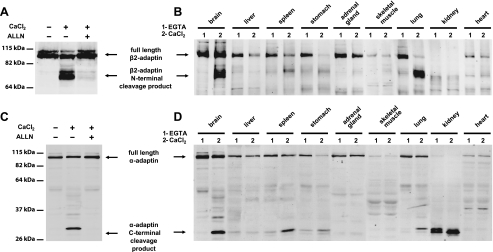
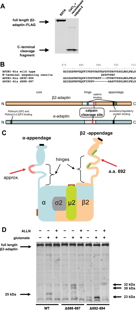
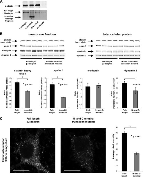
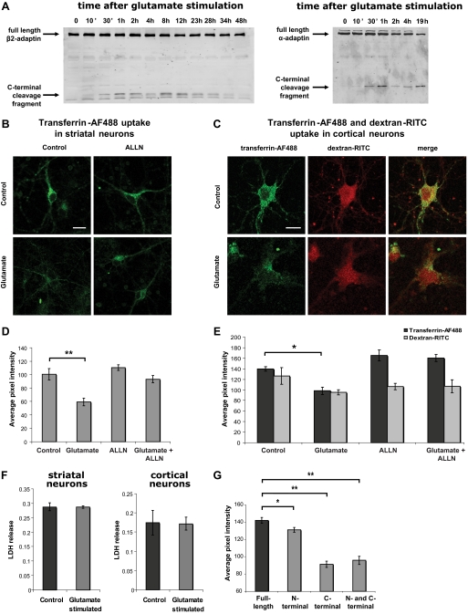
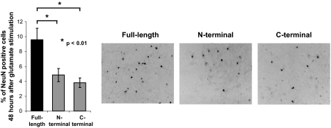
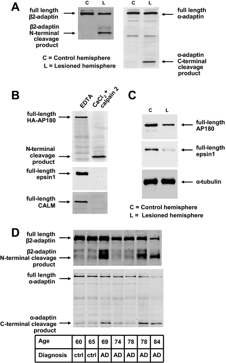
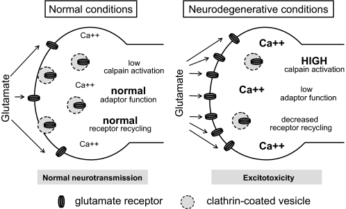
Clathrin-dependent endocytosis is mediated by a tightly regulated network of molecular interactions that provides essential protein-protein and protein-lipid binding activities. Here we report the hydrolysis of the alpha- and beta2-subunits of the tetrameric adaptor protein complex 2 by calpain. Calcium-dependent alpha- and beta2-adaptin hydrolysis was observed in several rat tissues, including brain and primary neuronal cultures. Neuronal alpha- and beta2-adaptin cleavage was inducible by glutamate stimulation and was accompanied by the decreased endocytosis of transferrin. Heterologous expression of truncated forms of the beta2-adaptin subunit significantly decreased the membrane recruitment of clathrin and inhibited clathrin-mediated receptor endocytosis. Moreover, the presence of truncated beta2-adaptin sensitized neurons to glutamate receptor-mediated excitotoxicity. Proteolysis of alpha- and beta2-adaptins, as well as the accessory clathrin adaptors epsin 1, adaptor protein 180, and the clathrin assembly lymphoid myeloid leukemia protein, was detected in brain tissues after experimentally induced ischemia and in cases of human Alzheimer disease. The present study further clarifies the central role of calpain in regulating clathrin-dependent endocytosis and provides evidence for a novel mechanism through which calpain activation may promote neurodegeneration: the sensitization of cells to glutamate-mediated excitotoxicity via the decreased internalization of surface receptors.


Figures

FIGURE 1.
FIGURE 1.
Cleavage of the β2- and α-subunits of the AP-2 complex in rat tissues. A, immunoblot analysis of rat brain homogenates with an antiserum that recognizes an N-terminal epitope of β2-adaptin. A novel molecular species consistent with a molecular mass of 75 kDa appears in rat brain extracts in the presence of CaCl2, the appearance of this band is blocked by the broad spectrum calpain inhibitor ALLN. B, immunoblot analysis of various rat tissue homogenates shows a calcium-dependent change in migration of the β2-adaptin immunoreactive species in brain, spleen, lung, and stomach. C, immunoblot analysis of rat brain homogenates with antiserum recognizing a C-terminal epitope of α-adaptin. A molecular species consistent with a molecular mass of 30 kDa appears in the rat brain protein extract when CaCl2 is present in the lysis buffer. This change in migration is blocked by the broad spectrum calpain inhibitor ALLN. D, immunoblot analysis of rat tissue homogenates using anti-C-terminal-α-adaptin antiserum shows a calcium-dependent change in migration of the immunoreactive species in samples of brain, spleen, lung, and stomach.
FIGURE 2.
FIGURE 2.
Cleavage of β2- and α-subunits of AP-2 in cultured striatal neurons. A and B, C-terminally 6×-histidine-tagged β2-adaptin-expressing primary neurons were stimulated with 100 μm glutamate for 15 min in the presence of 2.7 mm CaCl2 (or 3 mm EGTA) in combination with calpain 1 inhibitor PD 151746 (25 μm), calpain 2 inhibitor Z-LLY-FMK (10 μm), or NMDA-receptor antagonist MK801 (10 μm). Protein extracts were collected 1 h after the end of the glutamate application and subjected to immunoblotting with antibodies against the 6×-histidine tag (A) or α-adaptin (B). C, C-terminally 6×-histidine-tagged β2-adaptin expressed in HEK 293T cells is degraded in the same manner when exposed to purified calpain 1 and calpain 2, but not caspase 3. D, α-adaptin is degraded in the same manner when exposed to purified calpain 2.
FIGURE 3.
FIGURE 3.
Sites of calpain cleavage in AP-2. A, protein extracts from HEK 293T cells expressing β2-adaptin with C-terminal FLAG tag were treated with recombinant rat calpain 2 in presence of 2 mm CaCl2 and subjected to immunoblotting with an anti-FLAG tag antibody. B, text shows the alignment of amino acid sequences of wild-type β2-adaptin, protein sequencing results for the N terminus of its C-terminal calpain proteolytic fragment, and Δ692-694 and experimental Δ688-697 deletion mutants. The scheme represents the linearized domain structures of β2-adaptin (orange) and α-adaptin (blue). C, schematic representation of the heterotetrameric AP-2 complex, depicting binding sites for clathrin (bright green), clathrin accessory factors (black), and phosphatidylinositol phosphates (brown), as determined in previous studies (5-8). Red arrows indicate the approximate positions of calpain cleavage sites. D, cleavage of β2-adaptin at Ala-692 is abrogated by deletion mutations. Cultured striatal neurons were infected with lentiviral vectors encoding wild-type β2-adaptin, Δ692-694 or Δ688-697 deletion mutants, all with C-terminal 6×-histidine tags. Neurons were stimulated with 100 μm glutamate plus 2.7 mm CaCl2 for 15 min. Protein was extracted 1 h after the end of the glutamate stimulation, and the extracts were subjected to immunoblotting with an anti-6×-histidine antibody. The expected 25-kDa cleavage product of wild-type β2-adaptin is not observed with the deletion mutants (although both deletion mutants are partially cleaved by a calpain-like activity at alternate sites, as indicated by arrows at right); cleavage of both wild-type and mutant adaptors is blocked by the addition of the calpain inhibitor ALLN. These data are consistent with the presence of at least three potential calpain cleavage sites: the physiological one (described in Fig. 2) and two alternate sites that are utilized only after mutation of the endogenous proteolytic site. These findings are not unexpected given that calpain hydrolysis is governed largely by structural rather than primary sequence determinants (14).
FIGURE 4.
FIGURE 4.
Effects of β2-adaptin cleavage products on the membrane distribution of endocytosis-related proteins. A, an antibody against α-adaptin was used to immunoprecipitate AP-2-containing protein complexes from HEK 293T cells overexpressing wild-type β2-adaptin or β2-adaptin mutants comprising the N- or C-terminal products of calpain cleavage. Immunoblotting of the precipitates with an antibody against the N-terminal portion of β2-adaptin shows that the heterologously expressed N-terminal truncation mutant is present in the precipitated AP-2 complexes. B, membrane fractions and total lysates of HEK 293T cells transfected with plasmid vectors encoding wild-type β2-adaptin or N- and C-terminal truncation mutants were subjected to immunoblotting with antibodies against the clathrin heavy chain, dynamin 2, α-adaptin, and epsin 1. Fluorescence intensities of protein immunodetection in the cellular membrane fractions were quantified and normalized to the intensity of the corresponding bands in parallel samples of total cellular protein. Four samples per condition were analyzed. The amounts of pelletable clathrin heavy chain and epsin 1 were significantly lower in the cells overexpressing N- and C-terminal truncation mutants of rat β2-adaptin than in the cells overexpressing wild-type rat β2-adaptin. Dynamin 2 also showed a non-significant trend toward a decrease in the membrane fractions of the truncation mutant-expressing cells. The amount of membrane-associated α-adaptin was not changed by coexpression of N- and C-terminal β2-adaptin truncation mutants. C, primary cultured cortical neurons were transduced with a lentiviral encoding wild-type β2-adaptin or a combination of vectors encoding N- and C-terminal truncation mutants and immunostained with an antibody against clathrin heavy chain. For each cell, the immunofluorescence intensity was quantified in a 2-μm2 area at the visible limit of the cell border. β2-Adaptin truncation mutant-expressing cells demonstrated a significant decrease in clathrin immunostaining as compared with controls expressing wild-type β2-adaptin. Average pixel intensities were quantified from seven wild-type- and six truncation mutant-expressing cells. Bar = 10 μm.
FIGURE 5.
FIGURE 5.
Transferrin uptake in neurons after AP-2 subunit truncation or proteolysis. A, time course of β2- and α-adaptin cleavage after glutamate stimulation of striatal neurons. Striatal neurons were monitored for cleavage of C-terminal 6×-histidine-tagged β2-adaptin and wild-type α-adaptin cleavage after stimulation with 100 μm glutamate for 15 min in the presence of 2.7 mm CaCl2. Protein was extracted at the indicated time points after the end of the glutamate stimulation and the extracts were subjected to immunoblotting with anti-histidine tag or anti-α-adaptin antibodies, respectively. Cleavage of adaptins was observed within 30 min after neuronal stimulation. Cleavage, both β2- and α-adaptin, was also observed within 30 min in primary cultured cortical neurons (data not shown). B and D, uptake of Alexa Fluor 488-conjugated transferrin (transferrin-AF488) by striatal neurons was assessed 1 h after a 15-min stimulation with 100 μm glutamate as measured by confocal microscopy (representative images shown in the top panels). Transferrin-AF488 uptake was quantified from the confocal plane with the highest overall AF488 intensity by calculating the average pixel intensity for an area of ∼4 μm2 within the limits of the neuronal plasma membrane but excluding the neuronal nucleus. The following numbers of cells were analyzed for each condition: 210 control, 142 glutamate, 181 ALLN, 176 glutamate plus ALLN. Transferrin-AF488 uptake was significantly reduced in glutamate-treated samples, whereas the calpain inhibitor ALLN restored transferrin uptake to the basal level. Bar = 10 μm. C and E, decreased transferrin uptake was also observed in cortical neurons after glutamate-stimulated calpain activation and AP-2 proteolysis. Transferrin-AF488 and dextran-Rhodamine B isothiocyanate (dextran-RITC) uptake was quantified as for striatal cells with analysis of the following numbers of cells per group: 54 control, 54 glutamate, 52 ALLN, 61 glutamate plus ALLN. Uptake of the fluid-phase endocytosis indicator dextran-RITC (10 kDa) into the same compartment was not significantly altered by glutamate stimulation or ALLN treatment. Bar = 10 μm. F, no cell death was detected in striatal or cortical neurons within 1 h of glutamate stimulation, as analyzed by lactate dehydrogenase release into the culture medium. G, expression of N-terminal, C-terminal, or both truncation mutants of β2-adaptin via lentiviral vectors recapitulates the calpain-mediated effect on transferrin endocytosis. Transferrin uptake by neurons expressing mutants comprising the N- and C-terminal products of calpain cleavage were compared with the parallel lentiviral-induced expression of full-length wild-type β2-adaptin. The following numbers of cells were analyzed for each condition: 196 wild-type, 260 N-terminal alone, 79 C-terminal alone, and 105 N- and C-terminal combined. *, p < 0.01; **, p < 0.005.
FIGURE 6.
FIGURE 6.
β2-Adaptin truncation sensitizes neurons to excitotoxicity. Lentiviral expression of N- or C-terminal truncation mutants of β2-adaptin sensitizes striatal neurons to excitotoxicity, as compared with parallel expression of full-length wild-type β2-adaptin. The left bar graph shows % remaining NeuN-positive neuronal cells remaining 48 h after excitotoxic stimulation (compared with unstimulated cells). Right panels show representative fields of remaining NeuN-positive cells (appearing as dark profiles) in each of the three conditions.
FIGURE 7.
FIGURE 7.
Neurodegeneration-related cleavage of clathrin adaptors. A, immunoblotting shows that β2- and α-subunits of AP-2 are cleaved within 24 h after reperfusion in a rat transient cerebral ischemia model. B, accessory adaptors AP180, epsin 1, and CALM are also substrates of calpain 2. Lysates of HEK 293T cells overexpressing HA-tagged AP180 protein (top panel) or untransfected HEK293T cells (bottom two panels) were exposed to recombinant rat calpain 2 plus 2 mm CaCl2 and subjected to immunoblotting with antibodies against the HA tag, epsin 1, or CALM, respectively. C, immunoblotting with antibodies against AP180 and epsin 1 shows that these adaptors are proteolyzed in a rat transient cerebral ischemia model. D, immunoblots of human prefrontal cortex (Brodmann area 9) detect proteolysis of β2- and α-adaptins in three of five AD samples.
FIGURE 8.
FIGURE 8.
Model for calpain-driven sensitization of neuronal cells to excitotoxicity via decreased endocytosis. The left panel represents normal (non-degenerative) conditions, where calcium homeostasis is normal and calpain activity is low. The right panel represents a potential disease state where primary abnormalities of protein aggregation, energy metabolism, endoplasmic reticular stress, or dysregulated neurotransmission could potentially sensitize cells to excitatory neurotransmitters such as glutamate. In this case, calpain cleavage of endocytic proteins is envisaged to decrease excitatory receptor endocytosis, which would result in their increased presence on the neuronal surface and thereby mediate excitotoxic damage or death. Not shown is the possibility that an abnormal cell surface turnover of receptors could also result in their being abnormally trafficked to extrasynaptic sites, which could further sensitize them to negative effects of excitatory signals.

Similar articles

Cited by

References

    1. Kirchhausen, T. (2000) Annu. Rev. Biochem. 69 699-727 - PubMed
    1. Schmid, E. M., and McMahon, H. T. (2007) Nature 448 883-888 - PubMed
    1. Collins, B. M., McCoy, A. J., Kent, H. M., Evans, P. R., and Owen, D. J. (2002) Cell 109 523-535 - PubMed
    1. Robinson, M. S. (2004) Trends Cell Biol. 14 167-174 - PubMed
    1. Honing, S., Ricotta, D., Krauss, M., Spate, K., Spolaore, B., Motley, A., Robinson, M., Robinson, C., Haucke, V., and Owen, D. J. (2005) Mol. Cell 18 519-531 - PubMed

Publication types

MeSH terms

LinkOut - more resources