

Determinants of aquaporin-4 assembly in orthogonal arrays revealed by live-cell single-molecule fluorescence imaging
- PMID: 19240114
- PMCID: PMC2714425
- DOI: 10.1242/jcs.042341
Determinants of aquaporin-4 assembly in orthogonal arrays revealed by live-cell single-molecule fluorescence imaging
Abstract
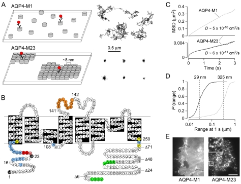
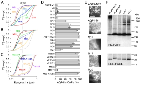
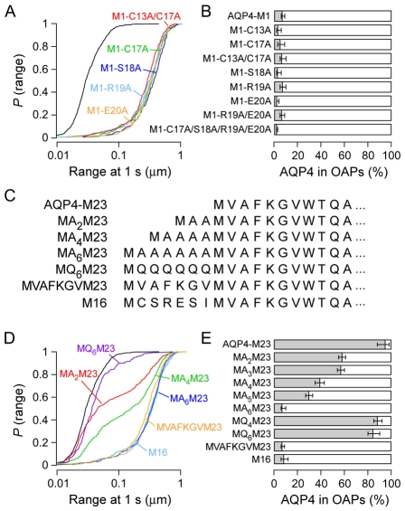
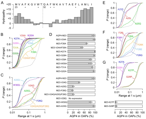
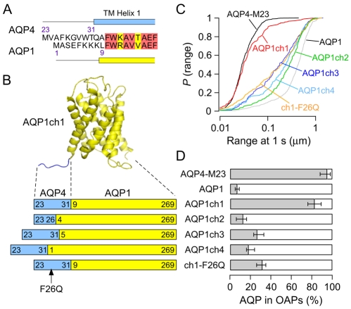
We investigated the molecular determinants of aquaporin-4 (AQP4) assembly in orthogonal arrays of particles (OAPs) by visualizing fluorescently labeled AQP4 mutants in cell membranes using quantum-dot single-particle tracking and total internal reflection fluorescence microscopy. The full-length ;long' (M1) form of AQP4 diffused freely in membranes and did not form OAPs, whereas the ;short' (M23) form of AQP4 formed OAPs and was nearly immobile. Analysis of AQP4 deletion mutants revealed progressive disruption of OAPs by the addition of three to seven residues at the AQP4-M23 N-terminus, with polyalanines as effective as native AQP4 fragments. OAPs disappeared upon downstream deletions of AQP4-M23, which, from analysis of point mutants, involves N-terminus interactions of residues Val24, Ala25 and Phe26. OAP formation was also prevented by introducing proline residues at sites just downstream from the hydrophobic N-terminus of AQP4-M23. AQP1, an AQP4 homolog that does not form OAPs, was induced to form OAPs upon replacement of its N-terminal domain with that of AQP4-M23. Our results indicate that OAP formation by AQP4-M23 is stabilized by hydrophobic intermolecular interactions involving N-terminus residues, and that absence of OAPs in AQP4-M1 results from non-selective blocking of this interaction by seven residues just upstream from Met23.
Figures
Similar articles
-
Reversible, temperature-dependent supramolecular assembly of aquaporin-4 orthogonal arrays in live cell membranes.Biophys J. 2009 Dec 2;97(11):3010-8. doi: 10.1016/j.bpj.2009.09.017. Biophys J. 2009. PMID: 19948131 Free PMC article.
-
Live cell analysis of aquaporin-4 m1/m23 interactions and regulated orthogonal array assembly in glial cells.J Biol Chem. 2009 Dec 18;284(51):35850-60. doi: 10.1074/jbc.M109.071670. J Biol Chem. 2009. PMID: 19843522 Free PMC article.
-
Live-cell imaging of aquaporin-4 diffusion and interactions in orthogonal arrays of particles.Neuroscience. 2010 Jul 28;168(4):892-902. doi: 10.1016/j.neuroscience.2009.08.034. Epub 2009 Aug 20. Neuroscience. 2010. PMID: 19699275 Free PMC article. Review.
-
Aquaporin-4 dynamics in orthogonal arrays in live cells visualized by quantum dot single particle tracking.Mol Biol Cell. 2008 Aug;19(8):3369-78. doi: 10.1091/mbc.e08-03-0322. Epub 2008 May 21. Mol Biol Cell. 2008. PMID: 18495865 Free PMC article.
-
Live-cell imaging of aquaporin-4 supramolecular assembly and diffusion.Methods Enzymol. 2012;504:341-54. doi: 10.1016/B978-0-12-391857-4.00017-3. Methods Enzymol. 2012. PMID: 22264543 Free PMC article. Review.
Cited by
-
Characterization of the binding pattern of human aquaporin-4 autoantibodies in patients with neuromyelitis optica spectrum disorders.J Neuroinflammation. 2016 Jul 1;13(1):176. doi: 10.1186/s12974-016-0642-3. J Neuroinflammation. 2016. PMID: 27371173 Free PMC article.
-
Model of aquaporin-4 supramolecular assembly in orthogonal arrays based on heterotetrameric association of M1-M23 isoforms.Biophys J. 2011 Jun 22;100(12):2936-45. doi: 10.1016/j.bpj.2011.05.012. Biophys J. 2011. PMID: 21689527 Free PMC article.
-
Aquaporin-4 in Neuromyelitis Optica Spectrum Disorders: A Target of Autoimmunity in the Central Nervous System.Biomolecules. 2022 Apr 17;12(4):591. doi: 10.3390/biom12040591. Biomolecules. 2022. PMID: 35454180 Free PMC article. Review.
-
The Water Transport System in Astrocytes-Aquaporins.Cells. 2022 Aug 18;11(16):2564. doi: 10.3390/cells11162564. Cells. 2022. PMID: 36010640 Free PMC article. Review.
-
Aquaporin Protein-Protein Interactions.Int J Mol Sci. 2017 Oct 27;18(11):2255. doi: 10.3390/ijms18112255. Int J Mol Sci. 2017. PMID: 29077056 Free PMC article. Review.
References
-
- Amiry-Moghaddam, M., Frydenlund, D. S. and Ottersen, O. P. (2004). Anchoring of aquaporin-4 in brain: molecular mechanisms and implications for the physiology and pathophysiology of water transport. Neuroscience 129, 999-1010. - PubMed
-
- Arluison, V., Seguin, J., Le Caer, J. P., Sturgis, J. N. and Robert, B. (2004). Hydrophobic pockets at the membrane interface: an original mechanism for membrane protein interactions. Biochemistry 43, 1276-1282. - PubMed
-
- de Groot, B. L., Engel, A. and Grubmuller, H. (2001). A refined structure of human aquaporin-1. FEBS Lett. 504, 206-211. - PubMed
Publication types
MeSH terms
Substances
Grants and funding
LinkOut - more resources
Full Text Sources
Other Literature Sources
