. 2009 Mar 15;122(Pt 6):849-58.
doi: 10.1242/jcs.041731. Epub 2009 Feb 24.

A novel role for SED1 (MFG-E8) in maintaining the integrity of the epididymal epithelium

Affiliations

A novel role for SED1 (MFG-E8) in maintaining the integrity of the epididymal epithelium

Adam S Raymond et al. J Cell Sci. .

Abstract

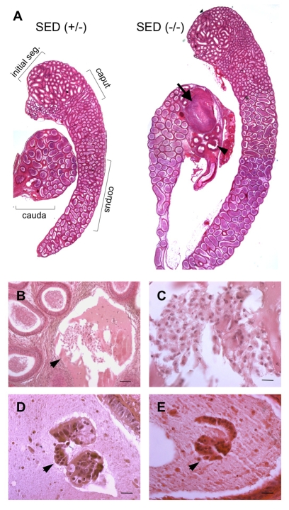
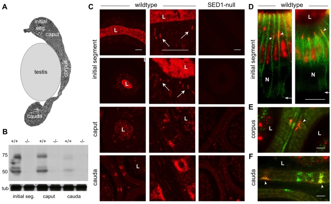
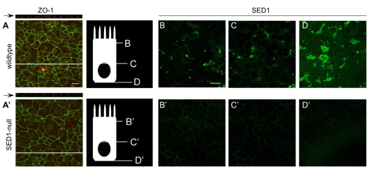
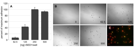
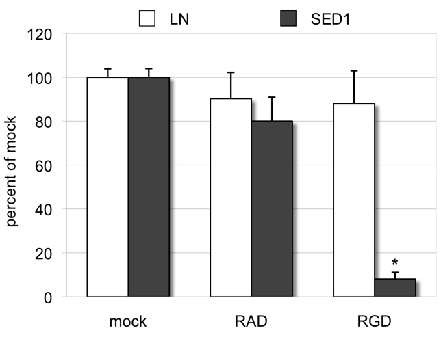
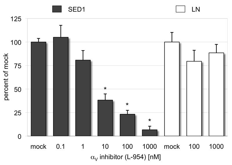
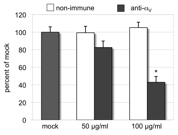
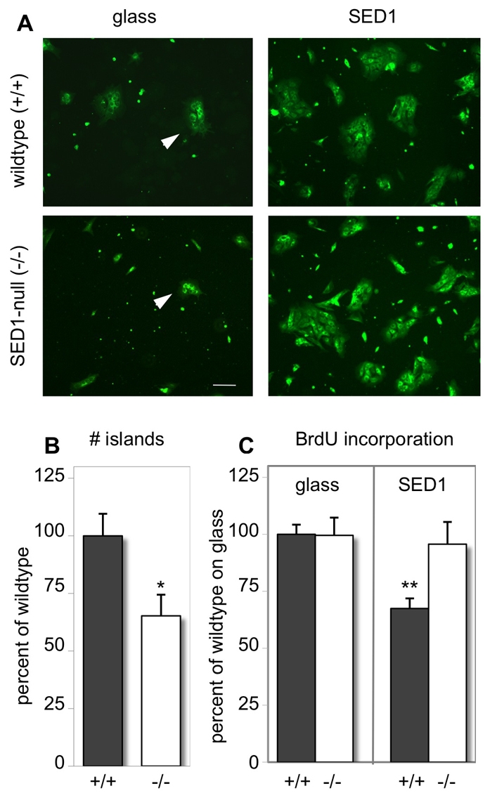
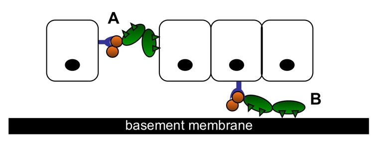
The epididymis is a highly convoluted tubule that connects the testis with the vas deferens, and in which mammalian sperm acquire the ability to fertilize eggs. The most proximal portion of the epididymis, or initial segment, secretes numerous factors that are critical for sperm maturation and storage. One such factor is SED1 (also known as MFG-E8) a bi-motif protein composed of two N-terminal EGF domains, the second of which contains an RGD motif, and two C-terminal discoidin domains (also known as F5/8 type C domains). Previous studies have reported that SED1 is secreted into the epididymal lumen, where it coats sperm and later facilitates sperm-egg binding. Herein, we report that SED1-null males also harbor unexpected epididymal pathologies, including detached epithelia and spermatic granulomas. We therefore examined whether SED1 has a tissue-intrinsic role in the epididymis, in addition to its role in sperm-egg adhesion. Improved fixation protocols revealed that SED1 is found in the basolateral domains of epididymal epithelial cells in vivo, and similarly, SED1 is secreted both apically and basally from polarized epididymal cells in vitro. The basolateral distribution of SED1 suggests that it may play a novel role in epididymal cell adhesion. Consistent with this, in vitro assays showed that SED1 supports epididymal cell adhesion via RGD binding to alphaV integrin receptors on epididymal epithelial cells. Finally, epididymal cells from SED1-null males showed reduced adhesion in vitro, a phenotype that can be rescued with exogenous SED1. These results suggest that SED1 facilitates epididymal cell adhesion, and that its loss leads to breakdown of the epididymal epithelium and consequent development of spermatic granulomas.


Figures

Fig. 1.
Fig. 1.
Epididymides from SED1-null males show epithelial breakdown. (A) Photomicrographs of paraffin-embedded epididymal tissue with the major functional regions identified. Sagittal sections from SED1 heterozygous (+/–) and null (–/–) littermates at 8 weeks of age stained with hematoxylin and eosin and imaged at identical magnification. The caudal segment of the SED1-null (–/–) epididymis contains a large spermatic granuloma (arrow) characterized by breakdown of the tubule and resulting in a bolus of sperm, fluid and immune cells. Occlusion of the tubule results in severe tubule swelling upstream of the lesion (compare corpus regions) and an absence of sperm in downstream tubules (arrowhead). (B-E) Other SED1-null pathologies not directly associated with the lesion site include detached, free-floating cells within the lumen suggestive of shed epithelium (arrowheads, B,D,E). (C) is an enlargement of (B). Scale bars: 50 μm (B), 20 μm (C-E).
Fig. 2.
Fig. 2.
SED1 localizes to both apical and basolateral domains of epididymal epithelial cells in vivo. (A) Composite photomicrograph of an epididymal sagittal-section positioned adjacent to a size-appropriate schematic of the testis. The initial segment, caput, corpus and cauda regions are identified. (B) Immunoblotting for SED1 in three epididymal regions; samples are loaded with equal protein. Immunoblotting of wild-type (+/+) tissue results in two bands consistent with the small and large isoforms of SED1. SED1-null (–/–) tissue produces background immunoreactivity. β-tubulin (tub) serves as a loading control. (C) In the initial segment, SED1 is found in punctate and often filamentous arrays along basolateral borders between adjacent epithelial cells (tissue perfused in 4% PFA). SED1 is also found in vesicles associated with the basal domain of nonprincipal cells, known as clear cells, in the caput, corpus and cauda segments (tissue submersion-fixed in Bouin's). SED1-null (–/–) tissue shows only background immunoreactivity. (D) Double-label immunofluorescence with E-cadherin (green) illustrates that many of the SED1 plagues (red) seen in the initial segment lie on the cell border (arrowheads). Two examples are shown. The base of the epithelium is designated by small arrows. (E,F) SED1-positive (red) clear cells of the caput and corpus regions fail to stain for the lysosomal marker, LAMP-1 (green), although clear cells in more distal regions (i.e. caput), are LAMP-1 positive, suggesting the protein is not destined for degradation in the proximal segments (caput, corpus) as it appears to be in the distal cauda. Images in panel C represent a merged stack of confocal Z-sections taken through the tissue, whereas those in panels D-F are merged epifluorescence photomicrographs. L, lumen; N, nucleus. Scale bars: 20 μm (C); 10 μm (D-F).
Fig. 3.
Fig. 3.
Polarized primary epididymal epithelial cells secrete SED1 both apically and basally. (A,A′) Immunofluorescence of the apical zonal occludin protein ZO-1 (green) and basal nuclei (red) in primary epididymal epithelial cells grown on Matrigel-coated transwell filters. X-Y projections and companion z-plane cross-sections (black arrow; white line indicates cross-section location) generated by confocal z-stack imaging show wild-type and SED1-null primary cells polarize and form apical tight junctions in culture. The cell diagram illustrates the approximate depth of confocal scans taken of polarized cultures immunostained with SED1 antisera. (B) In polarized cultures of wild-type cells, SED1 appears as punctate bodies in the apical domain of the cell consistent with its known apical secretion. (C) Little SED1 protein is found at the level of the nuclei; however immunoreactivity returns in the sub-nuclear basal domain (D). Examination reveals this signal is found in the same z-plane as the filter suggesting cells deposit SED1 onto the underlying substrate. (B′,C′,D′) SED1-null cultures assayed in parallel have no immunoreactivity. Scale bars: 20 μm.
Fig. 4.
Fig. 4.
Exogenous SED1 supports epididymal epithelial cell adhesion in a dose-dependent manner. (A) Increasing levels of SED1 lead to a concomitant increase in cell adhesion, reaching maximal values at 250 ng/well. Cells do not adhere to uncoated `mock' wells. (B) Representative photomicrographs of crystal violet-stained adherent cells on SED1 substrates. (C) Cytokeratin immunoreactivity confirms that the adherent cells are predominately epithelial. red, cytokeratin; green, nuclei (SYTO24). Error bars: s.d.
Fig. 5.
Fig. 5.
The addition of RGD peptides is sufficient to block the initial adhesion of epididymal epithelial cells to SED1. Cells preincubated with medium supplemented with 100 μM RGD or control (RAD) peptides were added to 30-minute adhesion assays on substrates coated with 500 ng/well SED1 or laminin. Data are expressed as a percentage of untreated (mock) cell adhesion to each substrate. RGD reduces adhesion to SED1 by 92% but has no effect on adhesion to laminin. RAD has a mild nonspecific effect on cell adhesion to both substrates. Error bars: s.e.m. *P<0.0001.
Fig. 6.
Fig. 6.
The αV integrin subunit is expressed in epididymal epithelial cells and localizes to focal plaques in cells adherent to SED1. (A) Immunoblotting for αV in the three epididymal regions; samples are loaded with equal protein. Immunoblotting of wild-type (+/+) and SED1-null (–/–) tissue under nonreducing conditions results in a single prominent band at 125 kDa, the reported molecular weight for αV under nonreduced conditions. Wild-type and SED1-null tissue express αV at similar levels. (B) Similarly, lysates of epithelial-enriched primary epididymal cells immunoprobed for αV exhibit a single 125 kDa band. β-tubulin serves as a loading control. (C) Immunostaining of cells cultured on SED1 reveals cytokeratin-positive epididymal epithelial cells (red) that express αV integrins (green) in punctate bundles arranged along the basal surface. (D) Desmin-positive (red) smooth muscle cells exhibit little or no αV immunoreactivity. Nonimmune (NI) stained cells produce background immunoreactivity. All images are from parallel experiments imaged under identical conditions. (E) Confocal micrographs reveal that cytokeratin-positive (inserts) epithelial cells cultured on SED1 localize αV to focal plaques along the lamellipodia (arrows). (F) Epithelial cells cultured on laminin also express αV; however, the immunoreactivity is not distributed in focal plaques as on SED1 substrates but remains perinuclear. Scale bars: 20 μm.
Fig. 7.
Fig. 7.
A small molecule inhibitor of αVβ3 and αVβ5 integrin heterodimers (L-954) selectively blocks epididymal cell adhesion to SED1. Increasing concentrations of L-594 were added to 30-minute adhesion assays, eliciting a dose-dependent decrease in adhesion to SED1 substrates (500 ng/well). The inhibitor has little effect on cell adhesion to laminin-coated substrates (500 ng/well). Data are expressed as a percentage of untreated (mock) cell adhesion to each substrate. As little as 1 nM of inhibitor is sufficient to reduce adhesion to SED1 by 19%, while 1000 nM inhibits adhesion by 93%. Error bars: s.e.m. *P<0.0001.
Fig. 8.
Fig. 8.
Function-blocking antibodies against αV integrin block epididymal cell adhesion to SED1. The addition of αV-blocking IgG (RMV-7) (50 μg/ml or 100 μg/ml) results in a dose-dependent reduction in epithelial cell adhesion to SED1 of 18% and 57%, respectively. Control IgG has no effect. Data are expressed as a percentage of untreated (mock) cell adhesion. Error bars: s.e.m. *P<0.0001.
Fig. 9.
Fig. 9.
Loss of SED1 leads to deficient cellular adhesion in vitro. Primary epithelial-enriched epididymal cells were isolated and cultured for 4 days on uncoated glass or SED1 substrates. Attached cells were pulsed with BrdU for 4 hours, fixed, and immunostained for BrdU. Epididymal `islands' (arrowheads) containing ≥4 nuclei were counted. (A) Epifluorescent imaging of SYTO24 counterstain illustrates a reduction in the number of SED1-null (–/–) epithelial islands relative to wild type (+/+). This phenotype is rescued when cells are cultured on SED1 substrates; under these conditions cells of both genotypes produce abundant islands. (B) Quantification shows that SED1-null (–/–) cells generate 35% fewer islands than wild-type (+/+) cells when cultured on glass substrates. (C) BrdU-positive nuclei associated with epithelial islands were counted and expressed as a percentage of total nuclei associated with islands. Data from multiple experiments were averaged and the wild-type (+/+) rate of proliferation was normalized to 100%. Although SED1-null (–/–) cells generate fewer islands than wild type, the rate of proliferation for the two genotypes is identical on glass substrates. Furthermore, the presence of SED1 substrates does not lead to increased proliferation relative to cells cultured on glass. Scale bar: 100 μm. Error bars: s.e.m. *P=0.01; **P<0.001.
Fig. 10.
Fig. 10.
Working model of SED1-dependent epididymal epithelial cell adhesion. The EFG domains (orange circles) and discoidin domains (green ovals) of SED1 are depicted bound to epididymal epithelial cells. The second EGF domain of SED1 contains a RGD motif and is proposed to serve as ligand for αVβ3 or αVβ5 integrins (blue receptor) expressed by the epithelial cells. Two models of adhesion are presented: (A) cell-cell adhesion and (B) cell-basement membrane adhesion. The discoidin domains are predicted to function by binding either (A) anionic phospholipids on the cell surface or (B) negatively charged residues within the basement membrane.

Similar articles

Cited by

References

    1. Abe, K., Takano, H. and Ito, T. (1983). Ultrastructure of the mouse epididymal duct with special reference to the regional differences of the principal cells. Arch. Histol. Jpn. 46, 51-68. - PubMed
    1. Abou-Haèila, A. and Fain-Maurel, M. A. (1984). Regional differences of the proximal part of mouse epididymis: morphological and histochemical characterization. Anat. Rec. 209, 197-208. - PubMed
    1. Andersen, M. H., Berglund, L., Rasmussen, J. T. and Petersen, T. E. (1997). Bovine PAS-6/7 binds alpha v beta 5 integrins and anionic phospholipids through two domains. Biochemistry 36, 5441-5446. - PubMed
    1. Andersen, M. H., Graversen, H., Fedosov, S. N., Petersen, T. E. and Rasmussen, J. T. (2000). Functional analyses of two cellular binding domains of bovine lactadherin. Biochemistry 39, 6200-6206. - PubMed
    1. Atabai, K., Fernandez, R., Huang, X., Ueki, I., Kline, A., Li, Y., Sadatmansoori, S., Smith-Steinhart, C., Zhu, W., Pytela, R. et al. (2005). Mfge8 is critical for mammary gland remodeling during involution. Mol. Biol. Cell 16, 5528-5537. - PMC - PubMed

Publication types

MeSH terms