. 2009 Feb 15;23(4):482-95.
doi: 10.1101/gad.1752109.

Regulation of SR protein phosphorylation and alternative splicing by modulating kinetic interactions of SRPK1 with molecular chaperones

Affiliations

Regulation of SR protein phosphorylation and alternative splicing by modulating kinetic interactions of SRPK1 with molecular chaperones

Xiang-Yang Zhong et al. Genes Dev. .

Abstract

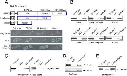
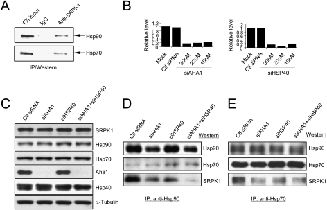
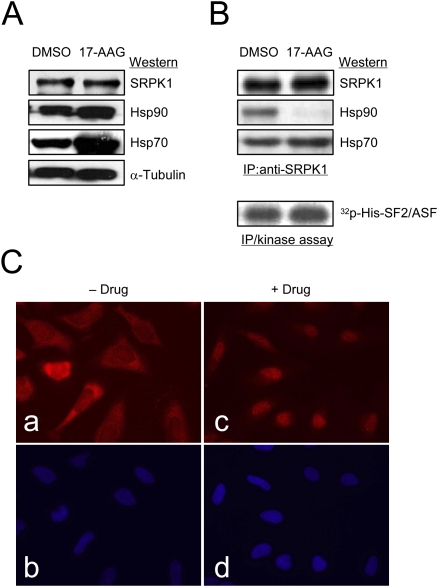
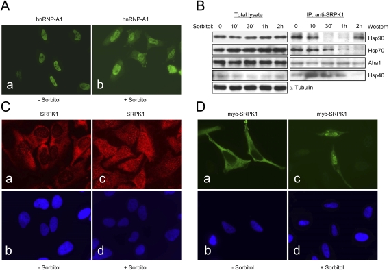
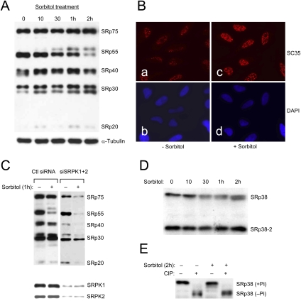
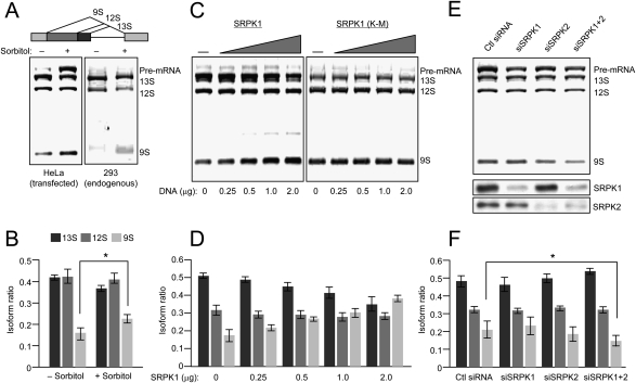
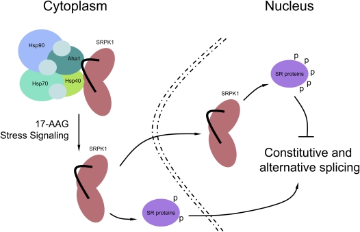
Phosphorylation is essential for the SR family of splicing factors/regulators to function in constitutive and regulated pre-mRNA splicing; yet both hypo- and hyperphosphorylation of SR proteins are known to inhibit splicing, indicating that SR protein phosphorylation must be tightly regulated in the cell. However, little is known how SR protein phosphorylation might be regulated during development or in response to specific signaling events. Here, we report that SRPK1, a ubiquitously expressed SR protein-specific kinase, directly binds to the cochaperones Hsp40/DNAjc8 and Aha1, which mediate dynamic interactions of the kinase with the major molecular chaperones Hsp70 and Hsp90 in mammalian cells. Inhibition of the Hsp90 ATPase activity induces dissociation of SRPK1 from the chaperone complexes, which can also be triggered by a stress signal (osmotic shock), resulting in translocation of the kinase from the cytoplasm to the nucleus, differential phosphorylation of SR proteins, and alteration of splice site selection. These findings connect the SRPK to the molecular chaperone system that has been implicated in numerous signal transduction pathways and provide mechanistic insights into complex regulation of SR protein phosphorylation and alternative splicing in response to developmental cues and cellular signaling.


Figures

Figure 1.
Figure 1.
Identification and validation of SRPK1-interacting proteins. (A) The bait constructs used in the two-hybrid screening. In full-length SRPK1, conserved kinase domains are split by a unique spacer sequence. The spacer was also constructed in the bait vector and used in a separate screen. The mutant SRPK1 deleted of the spacer (K1ΔSpacer) is toxic when stably expressed in yeast. This construct was only used for validation in transient assays. Pairwise two-hybrid interactions are indicated by both growth and the activity of the MEL1 reporter. (B) GST pull-down assay to confirm direct interactions between SRPK1 and specific chaperones in vitro. GST or GST fusion proteins as indicated were used to pull down recombinant SRPK1, K1ΔSpacer, or Spacer expressed and purified from bacteria. (C) GST or GST fusion proteins were also used to pull down SRPK1 from HeLa lysate. (D) Interaction of SRPK1 with Aha1 and Hsp40 in vivo. SRPK1 was immunoprecipitated from HeLa lysate followed by Western blotting analysis using anti-Aha1 and anti-pan-Hsp40 antibodies, the latter of which recognize multiple members of the Hsp40 family. (E) Specific interaction between SRPK1 and DNAjc8/Hsp40 in vivo. The HA-tagged DNAjc8/Hsp40 was transfected into HEK293 cells followed by anti-HA IP. SRPK1 in the complex was detected with anti-SRPK1. An empty vector was transfected and analyzed in parallel as a background control.
Figure 2.
Figure 2.
Cochaperone-mediated association of SRPK1 with major heat-shock proteins. (A) Detection of Hsp70 and Hsp90 in SRPK1 complexes. (B) siRNA-mediated knockdown of Aha1 and DNAjc8/Hsp40 transcripts, which was quantified by real time RT–PCR. Scrambled siRNA was tested as control. (C) Knockdown of the Aha1 protein in single and double siRNA-treated HeLa cells. We could not directly validate DNAjc8/Hsp40 knockdown because of the lack of a specific antibody. (D,E) Reduced association of SRPK1 with major chaperones in HeLa cells deleted of Aha1, DNAjc8/Hsp40, or both. The chaperone complex was immunoprecipitated by using anti-Hsp90 (D) and anti-Hsp70 (E). The amount of associated SRPK1 was dramatically reduced in the absence of both Aha1 (cochaperone for Hsp90) and DNAjc8/Hsp40 (cochaperone for Hsp70). A similar, but less reduction of SRPK1 in Hsp70-containing complexes was observed.
Figure 3.
Figure 3.
The Hsp90 ATPase activity is required for dynamic interaction of SRPK1 with the Hsp70/Hsp90 chaperone complex. (A) Total protein detected by Western blotting as indicated from HeLa cells treated with vehicle (DMSO) or with 17-demthoxygeldanamycin (17-AAG). Both Hsp70 and Hsp90 are known to be induced by the treatment with 17-AAG. (B) Release of SRPK1 from the Hsp90-containing complex upon the inhibition of the Hsp90 ATPase activity. The association of the kinase with Hsp70 seemed unaffected. SRPK1 released from the Hsp90-containg complex was equally, if not more, active as an SR protein kinase when tested by in vitro phosphorylation using the SR protein SF2/ASF as a substrate. (C) Induction of SRPK1 nuclear translocation by the Hsp90 ATPase inhibitor (Geldanamycin used in this experiment; similar results also obtained with 17-AAG.) (Panels a,c) Endogenous SRPK1 as detected by anti-SRPK1 before and after the drug treatment. (Panels b,d) Corresponding nuclei were stained by DAPI. The cellular distribution of SRPK1 was shifted from the cytoplasm to the nucleus in response to the drug treatment.
Figure 4.
Figure 4.
Nuclear translocation of SRPK1 in response to osmotic stress. (A) Osmotic stress-induced redistribution of hnRNP A1 from the nucleus to the cytoplasm as previously reported (van der Houven van Oordt et al. 2000). (B) Stress-induced release of SRPK1 from the chaperone complexes. (Left panel) Total proteins, including α-tubulin as a loading control, in the lysate from sorbitol-treated HeLa cells at different sorbitol treatment points were determined by Western blotting. Hsp70 was slightly induced by osmotic stress (Right panel). Disassociation of SRPK1 from the Hsp70/Hsp90 chaperones in response to osmotic stress was determined by anti-SRPK1 IP from equal amounts of cell lysate at different sorbitol treatment points followed by Western blotting analysis of key chaperone components as indicated. Interestingly, Aha1 remained associated with SRPK1 during the time course. Hsp90 appeared to return to SRPK1 after 2 h, indicating dynamic interactions of SRPK1 with the Hsp70/Hsp90 machinery. (C, panels a,c) Induction of SRPK1 nuclear translocation by osmotic stress. (Panels b,d) Corresponding nuclei were stained by DAPI. (D, panels a,c) Localization of myc-tagged SRPK1 in transfected HeLa cells before and after the sorbitol treatment. (Panels b,d) Nuclei in the fields were stained by DAPI. Nuclear translocation of the exogenously expressed SRPK1 was more dramatically induced relative to endogenous SRPK1.
Figure 5.
Figure 5.
Hyperphosphorylation of SR proteins in sorbitol-treated cells. (A) mAb104 analysis of the phosphorylation state of typical SR proteins in response to osmotic stress. The phosphoepitoses in SRp30 were elevated by the sorbitol treatment, while SRp55 was converted to a hyperphosphorylated form. In contrast, the phosphorylation level of SRp75 was modestly elevated, and that of SRp40 was little affected. α-tubulin was probed as a loading control (note that this control was also shown in Fig. 4B because the same bunch of cells was used in both experiments). (B) Immunostaining of SC35 in nuclear speckles in mock- and sorbitol-treated cells. Although immunostaining is not a quantitative assay, the phosphoepitope detected by anti-SC35 appears brighter in sorbitol-treated cells relative to that in mock-treated cells. (C) Role of SRPKs in mediating SR protein phosphorylation before and after the sorbitol treatment. HeLa cells were transfected with control siRNA or specific siRNAs against SRPK1 and SRPK2. (Bottom panel) The levels of both kinases were determined by Western blotting. Knockdown of both kinases results in reduced SR protein phosphorylation probed by using mAb104 without the sorbitol treatment. The level of SR protein phosphorylation went further down in SRPK-depleted cells treated in the presence of sorbitol, indicating a combined effect of diminished SRPK1 and SRPK2 and activated phosphatases. We loaded a similar amount of proteins in individual lanes using α-tubulin and total SF2/ASF as loading controls, which are not included in the panel. (D) In contrast to typical SR proteins, SRp38 became partially dephosphorylated as indicated by progressive increase in its gel mobility during the course of the sorbitol treatment. (E) Sorbitol-induced partial dephosphorylation of SRp38 was confirmed by CIP treatment.
Figure 6.
Figure 6.
The effect of sorbitol treatment on splice site selection. (A) Sorbitol induction of E1A alternative splicing in transfected HeLa (left panel) and 293T cells (right panel). The splicing pattern of the E1A reporter is illustrated above. (B) Quantification of the result from transfected HeLa cells; n = 3; (*) P < 0.05. (C) Impact of overexpressed wild-type and mutant SRPK1 on E1A alternative splicing. HeLa cells were transfected with increasing amounts of plasmids expressing wild-type (left panel) and mutant (right panel) SRPK1 along with the E1A reporter as indicated at the bottom. While wild-type SRPK1 induced the 9S isoform, the mutant SRPK1 lacked the effect, indicating the requirement for the kinase activity for the observed effect. (D) Quantification of the results from HeLa cells transfected with increasing concentrations of wild-type SRPK1 (n = 3). (E) Switch in splice site selection in response to siRNA-mediated knockdown of SRPK1 and SRPK2 in sorbitol-treated HeLa cells. The knockdown effects were verified by Western blotting as shown in the bottom two panels. (F) Quantification of the results in E; n = 3; (*) P < 0.05.
Figure 7.
Figure 7.
Model for the role of molecular chaperones in the regulation of SRPK1 nuclear translocation, SR protein phosphorylation, and pre-mRNA splicing.

References

    1. Aubol B.E., Chakrabarti S., Ngo J., Shaffer J., Nolen B., Fu X.D., Ghosh G., Adams J.A. Processive phosphorylation of alternative splicing factor/splicing factor 2. Proc. Natl. Acad. Sci. 2003;100:12601–12606. - PMC - PubMed
    1. Cao W., Jamison S.F., Garcia-Blanco M.A. Both phosphorylation and dephosphorylation of ASF/SF2 are required for pre-mRNA splicing in vitro. RNA. 1997;3:1456–1467. - PMC - PubMed
    1. Cardinali B., Cohen P.T., Lamond A.I. Protein phosphatase 1 can modulate alternative 5′ splice site selection in a HeLa splicing extract. FEBS Lett. 1994;352:276–280. - PubMed
    1. Colwill K., Pawson T., Andrews B., Prasad J., Manley J.L., Bell J.C., Duncan P.I. The Clk/Sty protein kinase phosphorylates SR splicing factors and regulates their intranuclear distribution. EMBO J. 1996;15:265–275. - PMC - PubMed
    1. Ding J.H., Zhong X.Y., Hagopian J.C., Cruz M.M., Ghosh G., Feramisco J., Adams J.A., Fu X.D. Regulated cellular partitioning of SR protein-specific kinases in mammalian cells. Mol. Biol. Cell. 2006;17:876–885. - PMC - PubMed

Publication types

MeSH terms

LinkOut - more resources