Relationships between brain activation and brain structure in normally developing children
- PMID: 19240138
- PMCID: PMC2758677
- DOI: 10.1093/cercor/bhp011
Relationships between brain activation and brain structure in normally developing children
Abstract
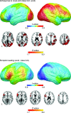
Dynamic changes in brain structure, activation, and cognitive abilities co-occur during development, but little is known about how changes in brain structure relate to changes in cognitive function or brain activity. By using cortical pattern matching techniques to correlate cortical gray matter thickness and functional brain activity over the entire brain surface in 24 typically developing children, we integrated structural and functional magnetic resonance imaging data with cognitive test scores to identify correlates of mature performance during orthographic processing. Fast-naming individuals activated the right fronto-parietal attention network in response to novel fonts more than slow-naming individuals, and increased activation of this network was correlated with more mature brain morphology in the same fronto-parietal region. These relationships remained even after effects of age or general cognitive ability were statistically controlled. These results localized cortical regions where mature morphology corresponds to mature patterns of activation, and may suggest a role for experience in mediating brain structure-activation relationships.
Figures


Similar articles
-
Individual differences in cognitive performance and brain structure in typically developing children.Dev Cogn Neurosci. 2015 Aug;14:1-7. doi: 10.1016/j.dcn.2015.05.003. Epub 2015 May 21. Dev Cogn Neurosci. 2015. PMID: 26046425 Free PMC article.
-
White matter growth as a mechanism of cognitive development in children.Neuroimage. 2006 Nov 15;33(3):936-46. doi: 10.1016/j.neuroimage.2006.07.024. Epub 2006 Sep 15. Neuroimage. 2006. PMID: 16978884
-
Brain structure and function correlates of general and social cognition.J Integr Neurosci. 2007 Mar;6(1):35-74. doi: 10.1142/s021963520700143x. J Integr Neurosci. 2007. PMID: 17472224
-
Imaging the developing brain: what have we learned about cognitive development?Trends Cogn Sci. 2005 Mar;9(3):104-10. doi: 10.1016/j.tics.2005.01.011. Trends Cogn Sci. 2005. PMID: 15737818 Review.
-
Neurobiological circuits regulating attention, cognitive control, motivation, and emotion: disruptions in neurodevelopmental psychiatric disorders.J Am Acad Child Adolesc Psychiatry. 2012 Apr;51(4):356-67. doi: 10.1016/j.jaac.2012.01.008. Epub 2012 Mar 3. J Am Acad Child Adolesc Psychiatry. 2012. PMID: 22449642 Review.
Cited by
-
Diffuse alterations in grey and white matter associated with cognitive impairment in Shwachman-Diamond syndrome: evidence from a multimodal approach.Neuroimage Clin. 2015 Feb 27;7:721-31. doi: 10.1016/j.nicl.2015.02.014. eCollection 2015. Neuroimage Clin. 2015. PMID: 25844324 Free PMC article.
-
Developmental changes in the structure of the social brain in late childhood and adolescence.Soc Cogn Affect Neurosci. 2014 Jan;9(1):123-31. doi: 10.1093/scan/nss113. Epub 2012 Oct 9. Soc Cogn Affect Neurosci. 2014. PMID: 23051898 Free PMC article.
-
Cortical anatomy in human X monosomy.Neuroimage. 2010 Feb 15;49(4):2915-23. doi: 10.1016/j.neuroimage.2009.11.057. Epub 2009 Dec 4. Neuroimage. 2010. PMID: 19948228 Free PMC article. Review.
-
Sex differences and structural brain maturation from childhood to early adulthood.Dev Cogn Neurosci. 2013 Jul;5:106-18. doi: 10.1016/j.dcn.2013.02.003. Epub 2013 Feb 24. Dev Cogn Neurosci. 2013. PMID: 23500670 Free PMC article.
-
A 3-arm randomized controlled trial on the effects of dance movement intervention and exercises on elderly with early dementia.BMC Geriatr. 2015 Oct 19;15:127. doi: 10.1186/s12877-015-0123-z. BMC Geriatr. 2015. PMID: 26481870 Free PMC article. Clinical Trial.
References
-
- Beckmann CF, Jenkinson M, Smith SM. General multilevel linear modeling for group analysis in FMRI. Neuroimage. 2003;20:1052–1063. - PubMed
-
- Brown TT, Lugar HM, Coalson RS, Miezin FM, Petersen SE, Schlaggar BL. Developmental changes in human cerebral functional organization for word generation. Cereb Cortex. 2005;15:275–290. - PubMed
-
- Colby CL, Goldberg ME. Space and attention in parietal cortex. Annu Rev Neurosci. 1999;22:319–349. - PubMed

