. 2009 Apr;37(7):2283-93.
doi: 10.1093/nar/gkp102. Epub 2009 Feb 24.

DNA uracil repair initiated by the archaeal ExoIII homologue Mth212 via direct strand incision

Affiliations

DNA uracil repair initiated by the archaeal ExoIII homologue Mth212 via direct strand incision

Lars Schomacher et al. Nucleic Acids Res. 2009 Apr.

Abstract

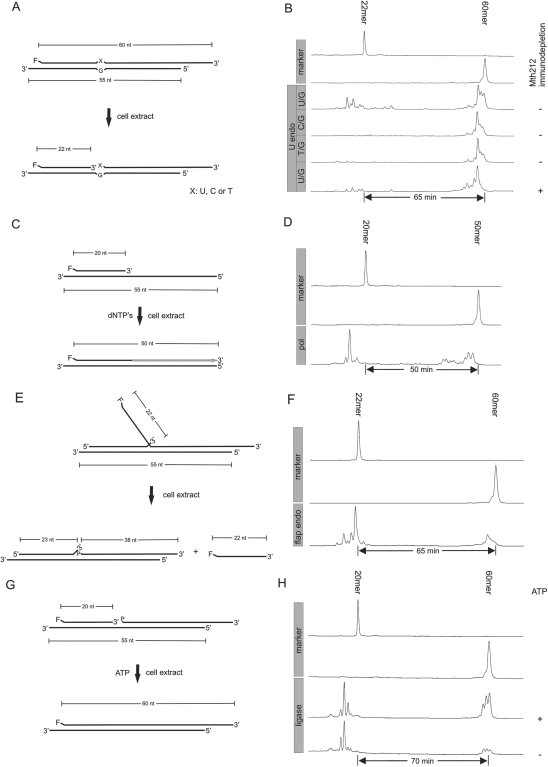
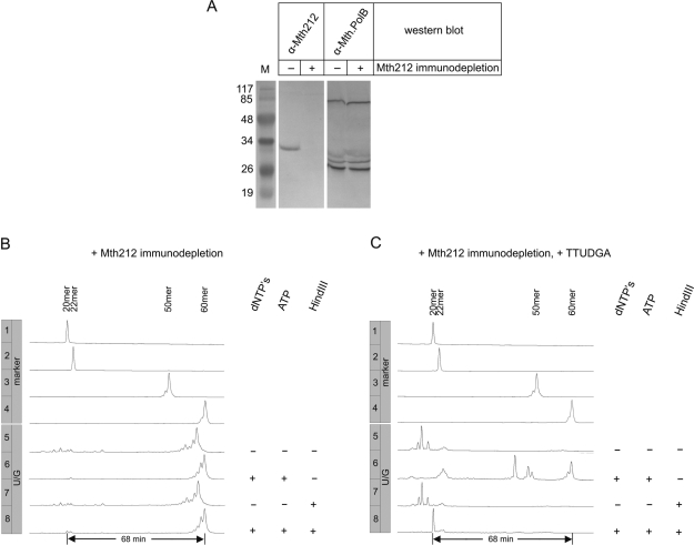
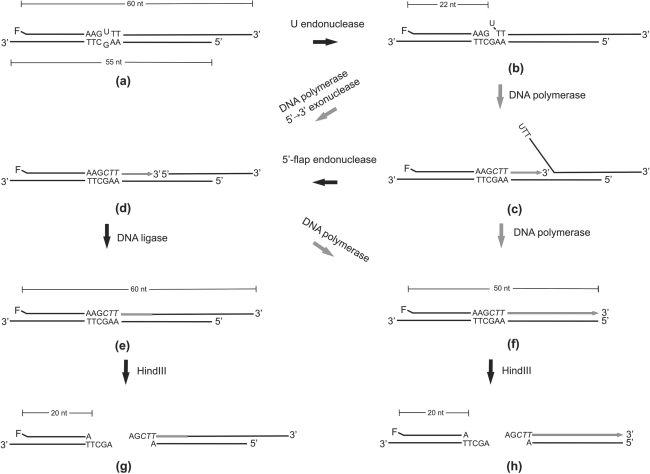
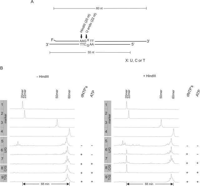
No genes for any of the known uracil DNA glycosylases of the UDG superfamily are present in the genome of Methanothermobacter thermautotrophicus DeltaH, making it difficult to imagine how DNA-U repair might be initiated in this organism. Recently, Mth212, the ExoIII homologue of M. thermautotrophicus DeltaH has been characterized as a DNA uridine endonuclease, which suggested the possibility of a novel endonucleolytic entry mechanism for DNA uracil repair. With no system of genetic experimentation available, the problem was approached biochemically. Assays of DNA uracil repair in vitro, promoted by crude cellular extracts, provide unequivocal confirmation that this mechanism does indeed operate in M. thermautotrophicus DeltaH.


Figures

Figure 1.
Figure 1.
Demonstration of enzymatic activities in cell extracts inferred to be required for uridine excision repair. (A), (C), (E), (G): Schematic drawings of substrates and expected reaction products (F: fluorescein moiety, nt: nucleotides). (B), (D), (F), (H): Trackings of fluorescence readouts as recorded by an ALF DNA sequencer. In lanes labeled ‘marker’, 5′-fluorescein conjugated oligonucleotides of known length were run; numbers of residues are indicated above respective peaks. Numbers of minutes refer to run time differences between marker oligonucleotides. (A), (B) DNA uridine endonuclease assay. (A) Three substrates differing with respect to the identity of residue ‘X’ (U, C or T), but otherwise being identical were tested. The left 3′-overhang is 5 nt in length, the right one 10 nt. (B) Reaction products of different substrates as indicated. α-Mth212 antibodies: ‘−’ or ‘+’ denote no treatment or pretreatment, respectively, of cell extract with α-Mth212 antibodies prior to reaction. (C), (D) DNA polymerase assay. C: Right 3′-overhang is as in (A). dNTPs: reaction was performed in presence of dNTP′s (20 µM each) in assay buffer. (E), (F) Flap endonuclease assay. (E) The flap branch point can move by one nucleotide yielding either a 22-nt or a 23-nt flap. Only the former structure is shown. 3′-overhangs are as in (A). ‘P’ denotes a 5′-phosphate residue. (G), (H): DNA ligase assay. G: 3′-overhangs are as in (A). ‘P’ denotes a 5′-phosphate residue. Ligase assay was carried out with or without ATP (5 mM) added to the assay buffer, as indicated in the right margin of (H). Conditions for all reactions were identical as described in detail in ‘Materials and Methods’ section.
Figure 4.
Figure 4.
DNA-U repair assays with extracts from M. thermautotrophicus ΔH immunochemically depleted for Mth212. (A) Western blot analysis of Mth212 immunodepletion. Untreated (‘−’) or Mth212 immunodepleted (‘+’) M. thermautotrophicus ΔH cell extracts were analyzed for the presence of Mth212 or Mth.PolB (control) by western blot as described in ‘Materials and methods’ section. M: Marker proteins (Fermentas) with corresponding molecular masses (×10−3). Calculated relative molecular weight for Mth212 is 30 350 and for the two subunits of Mth.PolB (14) 67 950 and 25 500. (B) Gel electrophoretic analysis of products after incubation of repair substrate (compare Figure 3) with cell extract immunochemically depleted for Mth212. dNTPs: presence (‘+’) or absence (‘−’) of dNTP's (20 µM each) in assay buffer. ATP: presence (‘+’) or absence (‘−’) of ATP (2 mM) in assay buffer. HindIII: no incubation (‘−’) or incubation (‘+’) of reaction products with HindIII. Lengths of 5′-fluorescein-labeled oligonucleotides are indicated above the respective peaks in lanes labeled ‘marker’. (C) Gel electrophoretic analysis of products after incubation of repair substrate with M. thermautotrophicus ΔH cell extract supplemented with 25 pmol of purified TTUDGA and then immunochemically depleted for Mth212. Other labels as in (B).
Figure 2.
Figure 2.
Outline of DNA-U repair assay. DNA polymerase reactions are represented by grey arrows, newly synthesized DNA is indicated by grey lines and letters in italics. The rationale underlying the assay is described in detail in the text.
Figure 3.
Figure 3.
DNA-U repair in cell extracts from M. thermautotrophicus ΔH. (A) Schematic representation of repair substrates (also compare figure 2). Arrows indicate cleavage points of U endonuclease (U endo, X = U) and HindIII (X = C, the C residue being provided with the substrate or introduced in the course of repair). (B) Gel electrophoretic analysis of reaction products after incubation of repair substrate with 50 µg cell extract from M. thermautotrophicus ΔH. dNTPs: presence (‘+’) or absence (‘−’) of dNTPs (20 µM each) in assay buffer. ATP: presence (‘+’) or absence (‘−’) of ATP (2 mM) in assay buffer. Sizes in nucleotides of 5′-fluorescein labeled oligonucleotides are given above the respective peaks in the lanes labeled ‘marker’. Left row of lanes (‘−HindIII’): without addition of HindIII. Right row of lanes (‘+ HindIII’): same reactions as in left panel, but after additional incubation with HindIII.

Similar articles

Cited by

References

    1. Lindahl T. Instability and decay of the primary structure of DNA. Nature. 1993;362:709–715. - PubMed
    1. Dianov G, Lindahl T. Reconstitution of the DNA base excision-repair pathway. Curr. Biol. 1994;4:1069–1076. - PubMed
    1. Kubota Y, Nash RA, Klungland A, Schar P, Barnes DE, Lindahl T. Reconstitution of DNA base excision-repair with purified human proteins: interaction between DNA polymerase beta and the XRCC1 protein. EMBO J. 1996;15:6662–6670. - PMC - PubMed
    1. Klungland A, Lindahl T. Second pathway for completion of human DNA base excision-repair: reconstitution with purified proteins and requirement for DNase IV (FEN1) EMBO J. 1997;16:3341–3348. - PMC - PubMed
    1. Sartori AA, Jiricny J. Enzymology of base excision repair in the hyperthermophilic archaeon Pyrobaculum aerophilum. J. Biol. Chem. 2003;278:24563–24576. - PubMed

MeSH terms