. 2009 Mar 17;106(11):4507-12.
doi: 10.1073/pnas.0900355106. Epub 2009 Feb 24.

Impaired maturation of dendritic spines without disorganization of cortical cell layers in mice lacking NRG1/ErbB signaling in the central nervous system

Affiliations

Impaired maturation of dendritic spines without disorganization of cortical cell layers in mice lacking NRG1/ErbB signaling in the central nervous system

Claudia S Barros et al. Proc Natl Acad Sci U S A. .

Abstract

Neuregulin-1 (NRG1) and its ErbB2/B4 receptors are encoded by candidate susceptibility genes for schizophrenia, yet the essential functions of NRG1 signaling in the CNS are still unclear. Using CRE/LOX technology, we have inactivated ErbB2/B4-mediated NRG1 signaling specifically in the CNS. In contrast to expectations, cell layers in the cerebral cortex, hippocampus, and cerebellum develop normally in the mutant mice. Instead, loss of ErbB2/B4 impairs dendritic spine maturation and perturbs interactions of postsynaptic scaffold proteins with glutamate receptors. Conversely, increased NRG1 levels promote spine maturation. ErbB2/B4-deficient mice show increased aggression and reduced prepulse inhibition. Treatment with the antipsychotic drug clozapine reverses the behavioral and spine defects. We conclude that ErbB2/B4-mediated NRG1 signaling modulates dendritic spine maturation, and that defects at glutamatergic synapses likely contribute to the behavioral abnormalities in ErbB2/B4-deficient mice.


Conflict of interest statement

The authors declare no conflict of interest.

Figures

Fig. 1.
Fig. 1.
Cortical development in ErbB2/B4-CNSko mice. (A) Immunopreciptiation of ErbB2 and ErbB4 from E14 and P0 brain extracts followed by Western blotting demonstrated an absence of ErbB2 and ErbB4 proteins; IP, immunoprecipitation. (B) Polymerase chain reaction (PCR) analysis of CRE-mediated recombination with DNA from P0 brains. In ErbB2/B4-CNSko mutants, the hGFAP-CRE transgene was detected, as well as a 170-kb (ErbB2) and a 507-kb (ErbB4) band indicating recombination of floxed alleles. (C) Brain size and morphology were unchanged in ErbB2/B4-CNSko mice at P249. (D) Sagittal brain sections at P38 stained with Nissl revealed no defects in cortical layers (roman numerals) and hippocampal structure. (E) Sagittal sections stained with NeuN (red) at P38, Cux1 at P256 (green), and Tbr1 at P249 (green). Nuclei were stained with Topro-3 (blue). (F) No differences were seen in NeuN, Cux1 or Tbr1-positive neurons in ErbB2/B4-CNSko mice (NeuN [cells/mm2]: WT 2665 ± 139, ErbB2/B4-CNSko 2630 ± 140, P > 0.05; Cux-1: WT 2015 ± 62, ErbB2/B4-CNSko 2052 ± 62, P > 0.05; Tbr1: WT 2257 ± 80, ErbB2/B4-CNSko 2192 ± 71, P > 0.05). (G) Coronal sections stained with the radial glia marker RC2 (green) and with antibodies to laminin-α2 (red) at embryonic ages E16 and E18. No difference in the gross morphology of radial glia was detected. (H) Immunoblotting for GFAP using brain extracts from P0 mice. Actin served as loading control. Scale bars, 50 μm
Fig. 2.
Fig. 2.
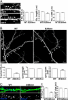
Dendritic spine loss. (A, B) CA1 pyramidal neurons at P26 stained with Golgi. (A) Dendrites of CA1 neurons. (B) Spines (arrowheads) along apical dendrites of CA1 neurons. (C) Spine density along apical dendrites was reduced in ErbB2/B4-CNSko mice (WT: 16.82 ± 0.49 spines/10 μm, n = 1367 spines from 26 neurons; ErbB2/B4-CNSko: 12.09 ± 0.66 spines/10 μm, n = 983 spines from 24 neurons; ***P < 0.001). (D, E) Pyramidal neurons in the prefrontal cortex stained by Golgi. (D) Cell bodies (arrowheads) and dendrites of pyramidal neurons. (E) Spine density along dendrites was reduced in ErbB2/B4-CNSko mice (WT: 9.29 ± 0.5 spines/10 μm, n = 483 spines from 20 neurons; ErbB2/B4-CNSko: 7.82 ± 0.44 spines/10 μm, n = 427 spines from 20 neurons; *P < 0.05). Scale bars, (A), 50 μm; (D), 100 μm; (B and E), 10 μm.
Fig. 3.
Fig. 3.
Spine development in cultured neurons. (A) Hippocampal neurons transfected with eGFP, cultured for 11 days (DIV11) and imaged to reveal filopodia (arrows). Filopodial density and width were unaltered in mutants, but length was reduced (density [filopodia/10 μm]: WT 5.85 ± 0.31, ErbB2/B4-CNSko 5.88 ± 0.37, P > 0.05; width [μm]: WT 0.25 ± 0.01, ErbB2/B4-CNSko 0.25 ± 0.01, P > 0.05; length [μm]: WT 2.35 ± 0.08, ErbB2/B4-CNSko 1.86 ± 0.1, ***P < 0.001; n = 650 filopodia from 14 WT neurons, n = 530 filopodia from 12 ErbB2/B4-CNSko neurons). (B) Hippocampal neurons were imaged at DIV21 to reveal spines (arrowheads). Spine density and width were reduced in neurons from mutants. No difference was detected in spine length: (density [spines/10 μm]: WT 8.92 ± 0.25, ErbB2/B4-CNSko 4.09 ± 0.37, ***P < 0.001; width [μm]: WT 0.6 ± 0.01, ErbB2/B4-CNSko 0.5 ± 0.01, ***P < 0.001; length [μm]: WT 1.3 ± 0.01, ErbB2/B4-CNSko 1.27 ± 0.04, P > 0.05; n = 442 spines from eight ErbB2/B4-CNSko neurons, n = 2852 spines from 34 WT neurons). (C) Immunostaining with VGLUT (blue) in hippocampal neurons transfected with eGFP (green). Blue channel is shown separately in gray. The density of VGLUT clusters was reduced in ErbB2/B4-CNSko neurons. No difference was observed in cluster size: (density [clusters/10 μm]: WT 7.23 ± 0.4, n = 509 clusters from 10 neurons; ErbB2/B4-CNSko 3.84 ± 0.22, n = 295 clusters from 10 neurons, ***P < 0.001; (size [pixels]: WT 52.13 ± 3.06 pixels, ErbB2/B4-CNSko 49.12 ± 2.23 pixels, P > 0.05). Scale bars, 10 μm.
Fig. 4.
Fig. 4.
Exogenous rNRG1 accelerates dendritic spine maturation. (A) WT hippocampal neurons at DIV11 transfected with eGFP (green) were treated with rNRG1 or vehicle (veh). In the presence of rNRG1, the density and width of filopodia-like protrusions (arrows) increased, but not their length: (density [protusions/10 μm]: WT+rNRG1 11.3 ± 0.53, WT+veh 9.71 ± 0.37, *P < 0.05; width μm]: WT+rNRG1 0.33 ± 0.01, WT+veh 0.28 ± 0.01, ***P < 0.001; length μm]: WT+rNRG1 1.36 ± 0.04, WT+veh 1.39 ± 0.06, P > 0.05; n = 768 protrusion from eight WT+rNRG1 neurons, n = 536 protrusions from 8 WT+veh neurons). (B) WT hippocampal neurons at DIV21 treated with rNRG1 or vehicle. The density and width of spines (arrowheads) was increased by rNRG1. Spine length was unaffected (density [spines/10 μm]: WT+rNRG1 16.2 ± 0.63, WT+veh 13.4 ± 0.57, **P < 0.01; width, μm]: WT+rNRG1 0.44 ± 0.01, WT+veh 0.39 ± 0.01, ***P < 0.001; length, μm]: WT+rNRG1 0.92 ± 0.01, WT+veh 0.88 ± 0.01, P > 0.05; n = 1142 spines from 9 WT+rNRG1 neurons, n = 861 spines from nine WT+veh neurons). (C) Immunostaining for VGLUT (blue) in DIV21 WT hippocampal neurons treated with rNRG1 or vehicle. Blue channel is shown separately in gray. In the presence of rNRG, VGLUT cluster density and size was increased (density [clusters/10 μm]: WT+rNRG1 8.31 ± 0.34, n = 851 clusters from 18 neurons; WT+veh 7.05 ± 0.35, n = 587 clusters from 13 neurons, *P < 0.05; size, pixels: WT+rNRG1 71.46 ± 3.98, WT+veh 52.68 ± 2.76, ***P < 0.001). Scale bars, 10 μm.
Fig. 5.
Fig. 5.
Loss of ErbB2/B4 affects interactions between NMDAr and PSD-95. (A) Hippocampal neurons from WT and ErbB2/B4-CNSko mice transfected with eGFP (green) were stained at DIV21 with antibodies to PSD-95 (red) and NMDAr subunit NR1 (blue). Higher magnifications with red and blue channels are also shown separately in gray. (B) The density of PSD-95 and NR1 clusters was reduced in mutants. NR1 density (clusters/10 μm]: WT 6.92 ± 0.45, n = 465 clusters from 10 neurons; ErbB2/B4-CNSko 4.17 ± 0.43, n = 353 clusters from 11 neurons, ***P < 0.001; PSD-95 density (clusters/10 μm): WT 6.9 ± 0.52, n = 464 clusters from 10 neurons; ErbB2/B4-CNSko 4.19 ± 0.36, n = 347 clusters from 11 neurons, ***P < 0.001. (C) In ErbB2/B4-CNSko neurons the percentage of co-localization of NR1 and PSD-95 clusters was reduced (%): WT 23.25 ± 0.85, ErbB2/B4-CNSko 16.88 ± 1.17, ***P < 0.001). (D) Co-immunoprecipitation of NR1, NR2A or NR2B with PSD-95 revealed a 64.8 ± 8.1% (*P < 0.05), 34.5 ± 2.5% (**P < 0.01), and 30.2 ± 5.7% (**P < 0.01) decrease of protein interactions in mutants. Reverse co-immunoprecipitation of PSD-95 with NR1 also showed a reduction of 18.2 ± 1.7% (**P < 0.01). For each co-immunoprecipitation assay, the amount of immunoprecipitated protein revealed by immunoblotting is also presented. Negative controls for co-immunoprecipitation assays are shown in Fig. S7C. Scale bars, 10 μm.
Fig. 6.
Fig. 6.
Behavioral abnormalities in ErbB2/B4-CNSko mice and spine loss are attenuated by clozapine treatment. (A, B) In open-field test, ErbB2/B4-CNSko mice showed no difference in distance traveled compared with WT (A; WT 2418 ± 500 cm, n = 17 animals; ErbB2/B4-CNSko 2218 ± 374 cm, n = 15 animals; P > 0.05), but remained longer in the central zone of the open field (B; WT 17 ± 2% of time, ErbB2/B4-CNSko 27% ± 4% of time, *P < 0.05). (C) Resident intruder assay showed that ErbB2/B4-CNSko mice engaged for longer time in aggressive behavior (biting, kicking, wrestling) compared with WT (WT 29.5 ± 9.3 seconds, n = 7 animals; ErbB2/B4-CNSko 122.8 ± 41.6 seconds, n = 5 animals; *P < 0.05). Clozapine (cloz) treatment decreased aggression in ErbB2/B4-CNSko mice to WT levels (ErbB2/B4-CNSko+cloz: 6.37 ± 5.99 seconds; WT without clozapine: 29.5 ± 9.3 seconds, P > 0.05), without affecting basal aggression in WT (WT+clozapine: 43.3 ± 11.2 seconds; WT without clozapine: 29.5 ± 9.3 seconds, P > 0.05). (D) In a PPI test, ErbB2/B4-CNSko mice showed reduced PPI compared with WT (WT 78.6% ± 4.3% PPI, n = 7 animals; ErbB2/B4-CNSko 57.4% ± 8.4% PPI, n = 5 animals; *P < 0.05). This difference was abolished after clozapine treatment (WT 80.7% ± 11.6% PPI, ErbB2/B4-CNSko 77.7% ± 6.2% PPI; P > 0.05). (E) Hippocampal neurons from WT and ErbB2/B4-CNSko mice at DIV21 treated with clozapine or vehicle (veh). In neurons from WT mice, clozapine treatment resulted in an increase in spine density, but spine width and length were unaffected: (density, spines/10 μm: WT+veh 10.56 ± 0.46, WT+cloz 12.7 ± 0.37, ***P < 0.001; (width, μm): WT+veh 0.51 ± 0.01, WT+cloz 0.51 ± 0.01, P > 0.05; (length, μm): WT + veh 1.13 ± 0.03, WT + cloz 1.08 ± 0.02, P > 0.05; n = 641 spines from 11 WT+veh neurons, n = 1058 spines from 13 WT+cloz neurons). In neurons from ErbB2/B4-CNSko mice, clozapine increased spine density, width, and length: density, spines/10 μm): ErbB2/B4-CNSko+veh 5.88 ± 0.35, ErbB2/B4-CNSko+cloz 8.83 ± 0.29, ***P < 0.001 (width, μm): ErbB2/B4-CNSko+veh 0.42 ± 0.01, ErbB2/B4-CNSko+cloz 0.5 ± 0.01, ***P < 0.001 (length, μm): ErbB2/B4-CNSko+veh 1.06 ± 0.03, ErbB2/B4-CNSko+cloz 1.2 ± 0.03, **P < 0.01; n = 518 spines from 13 ErbB2/B4-CNSko+veh neurons, n = 678 spines from 11 ErbB2/B4-CNSko+cloz neurons. Scale bars, 10 μm

Similar articles

Cited by

References

    1. Mei L, Xiong WC. Neuregulin 1 in neural development, synaptic plasticity and schizophrenia. Nat Rev Neurosci. 2008;9:437–452. - PMC - PubMed
    1. Benzel I, et al. Interactions among genes in the ErbB-Neuregulin signalling network are associated with increased susceptibility to schizophrenia. Behav Brain Funct. 2007;3:31–41. - PMC - PubMed
    1. Gassmann M, et al. Aberrant neural and cardiac development in mice lacking the ErbB4 neuregulin receptor. Nature. 1995;378:390–394. - PubMed
    1. Lee KF, et al. Requirement for neuregulin receptor erbB2 in neural and cardiac development. Nature. 1995;378:394–398. - PubMed
    1. Meyer D, Birchmeier C. Multiple essential functions of neuregulin in development. Nature. 1995;378:386–390. - PubMed

Publication types