

Determination of X-ray flux using silicon pin diodes
- PMID: 19240326
- PMCID: PMC2651761
- DOI: 10.1107/S0909049508040429
Determination of X-ray flux using silicon pin diodes
Abstract
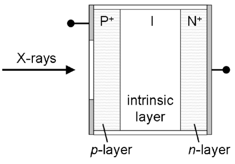
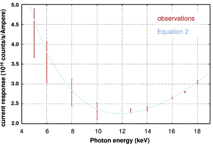
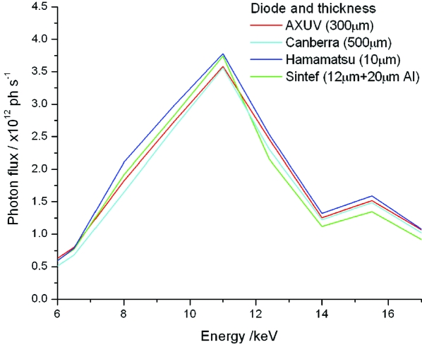
Accurate measurement of photon flux from an X-ray source, a parameter required to calculate the dose absorbed by the sample, is not yet routinely available at macromolecular crystallography beamlines. The development of a model for determining the photon flux incident on pin diodes is described here, and has been tested on the macromolecular crystallography beamlines at both the Swiss Light Source, Villigen, Switzerland, and the Advanced Light Source, Berkeley, USA, at energies between 4 and 18 keV. These experiments have shown that a simple model based on energy deposition in silicon is sufficient for determining the flux incident on high-quality silicon pin diodes. The derivation and validation of this model is presented, and a web-based tool for the use of the macromolecular crystallography and wider synchrotron community is introduced.
Figures
Comment in
-
Comments on Determination of X-ray flux using silicon pin diodes by R. L. Owen et al. (2009). J. Synchrotron Rad. 16, 143-151.J Synchrotron Radiat. 2009 Sep;16(Pt 5):690; author reply 690-1. doi: 10.1107/S0909049509023887. Epub 2009 Jul 2. J Synchrotron Radiat. 2009. PMID: 19713645 No abstract available.
References
-
- Alig, R. C. & Bloom, S. (1975). Phys. Rev. Lett.35, 1522–1525.
-
- Alkire, R. W. & Rotella, F. J. (1997). J. Appl. Cryst.30, 327–332.
-
- Bourenkov, G. P. & Popov, A. N. (2006). Acta Cryst. D62, 58–64. - PubMed
-
- Cho, T., Takahashi, E., Hirata, M., Yamaguchi, N., Teraji, T., Matsuda, K., Takeuchi, A., Kohagura, J., Yatsu, K., Tamano, T., Kondoh, T., Aoki, S., Zhang, X. W., Maezawa, H. & Miyoshi, S. (1992). Phys. Rev. A, 46, 3024–3027. - PubMed
-
- Cole, A. (1969). Radiat. Res.38, 7–33. - PubMed
Publication types
MeSH terms
Substances
Grants and funding
LinkOut - more resources
Full Text Sources
Other Literature Sources
