. 2009 Jun;17(6):1003-11.
doi: 10.1038/mt.2009.22. Epub 2009 Feb 24.

High mobility group box2 promoter-controlled suicide gene expression enables targeted glioblastoma treatment

Affiliations

High mobility group box2 promoter-controlled suicide gene expression enables targeted glioblastoma treatment

Poonam Balani et al. Mol Ther. 2009 Jun.

Abstract

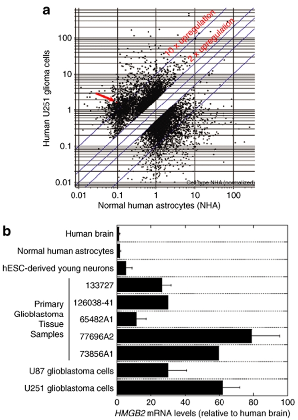
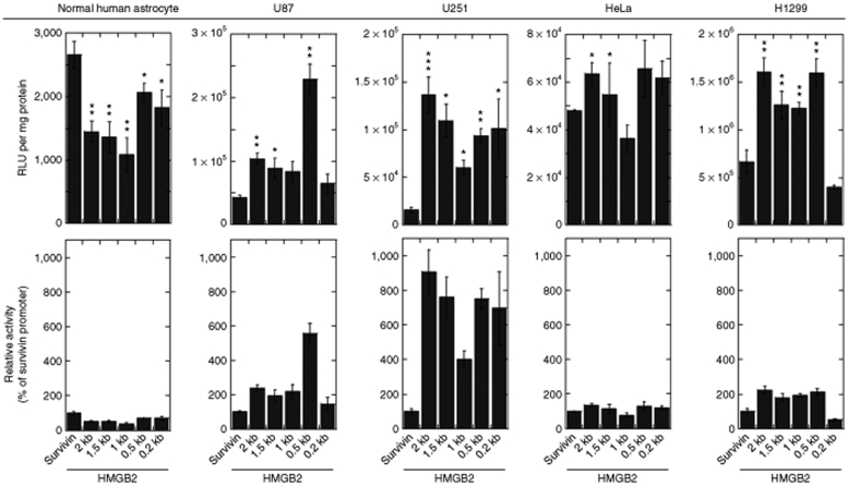
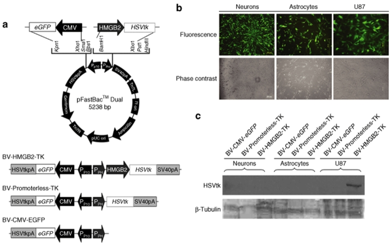
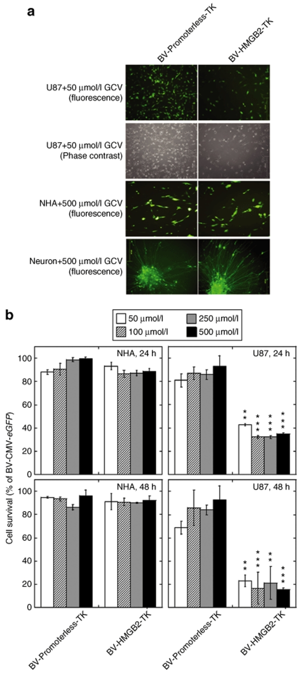
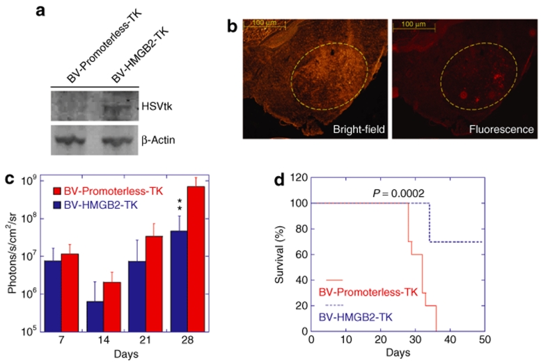
Achievement of specific tumor cell targeting remains a challenge for glioma gene therapy. We observed that the human high mobility group box2 (HMGB2) gene had a low level of expression in normal human brain tissues, but was significantly upregulated in glioblastoma tissues. With progressive truncation of a 5'-upstream sequence of the HMGB2 gene, we identified a 0.5-kb fragment displaying a high transcriptional activity in glioblastoma cells, but a low activity in normal brain cells. To test the feasibility of using the HMGB2 promoter sequence in targeted cancer therapy, we constructed a baculoviral vector expressing the herpes simplex virus thymidine kinase (HSVtk) gene driven by the HMGB2 promoter. Transduction with the viral vector induced cell death in glioblastoma cell lines in the presence of ganciclovir (GCV), but did not affect the survival of human astrocytes and neurons. In a mouse xenograft model, intratumor injection of the baculoviral vector suppressed the growth of human glioblastoma cells and prolonged the survival of tumor-bearing mice. Our results suggest that the novel 5' sequence of HMGB2 gene has a potential to be used as an efficient, tumor-selective promoter in targeted vectors for glioblastoma gene therapy.


Figures

<b>Figure 1</b>
Figure 1
Expression of HMGB2 mRNA in brain tissues and cells. (a) DNA microarray analysis of HMGB2 mRNA levels in U251 glioblastoma cells and normal human astrocytes. Three replicate Affimetrix HG_U133A_Plus_2 GeneChip arrays were used for each type of cells. Microarray results were analyzed using GeneSpring GX 7.3. The arrow indicates HMGB2 mRNA expression levels, which were low in normal human astrocytes, but more than tenfold upregulated in U251 glioblastoma cells. (b) Real-time PCR quantification of HMGB2 mRNA expression. Normal human brain tissue, primary glioblastoma tissues, normal human astrocytes and neurons, and U87 and U251 glioblastoma cell lines were used. hESC-derived young neurons were collected 1 week after neuronal differentiation of neural stem cells generated from hES1 human embryonic stem cells. The values represent the fold change in HMGB2 expression, normalized to β-actin mRNA levels and relative to HMGB2 expression in the normal brain tissue (means ± SD, n = 2–6). HMGB2, human high mobility group box2; mRNA, messenger RNA; hESC, human embryonic stem cells.
<b>Figure 2</b>
Figure 2
HMGB2 promoter activity in normal and tumor cells. Normal human astrocytes, U87 and U251 human glioma cell lines, HeLa human cervical cancer cell line and H1299 human lung cancer cell line were transfected with plasmid expression vectors accommodating a luciferase gene under the control of a HMGB2 promoter construct. A plasmid vector with a survivin promoter was included for comparison. Cells were lysed in a reporter lysis buffer 24 hours post transfection and promoter activity was measured using a luciferase assay system. Data are presented as relative light units per mg protein (upper panels) and as HMGB2 promoter activity relative to survivin promoter activity (lower panels). All data sets are a compilation of at least three independent experiments and expressed as mean ± SD. *P < 0.05, **P < 0.01, ***P < 0.001 vs. the survivin group by Student's t-test. HMGB2, human high mobility group box2.
<b>Figure 3</b>
Figure 3
HSVtk suicide gene expression driven by a HMGB2 promoter placed into a baculoviral vector. (a) Schematic representation of baculoviral constructs tested in this study. pFastBac Dual was used as the backbone for baculoviral vector construction. CMV-driven eGFP expression is used to monitor transduction efficiency and BV-Promoterless-TK is used as a negative control for HSVtk. (b) Baculoviral transduction in human neural cells and glioma cells. Human neurons derived from hES1 neural stem cells (Neurons) were transduced with BV-Promoterless-TK at an MOI 100. Normal human astrocytes (Astrocytes) and U87 glioblastoma cells (U87) were transduced BV-Promoterless-TK at an MOI 10. Fluorescence and phase contrast images taken 24 hours after transduction are shown. (c) HSVtk protein expression in vitro after baculoviral transduction. Cell lysates were prepared 24 hours after transduction. Ten µg of total protein from the cell lysates was used for western blotting. CMV, human cytomegalovirus immediate-early gene enhancer/promoter; eGFP, an enhanced green fluorescent protein gene; HMGB2, a 0.5-kb human high mobility group box 2 gene promoter; HSVtk, herpes simplex virus thymidine kinase gene; HSVtkpA, HSVtk polyadenylation signal; PPH, polyhedrin insect cell promoter; PP10, Insect cell p10 promoter; SV40pA, SV40 polyadenylation signal.
<b>Figure 4</b>
Figure 4
Selective cytotoxic effects of HMGB2 promoter-driven suicide gene expression. (a) Cellular effects as visualized under a microscope. Cells were transduced for 24 hours with either BV-HMGB2-TK or BV-Promoterless-TK, at an MOI 10 for astrocytes and U87 cells and at an MOI 100 for neurons, followed by treatment with the prodrug ganciclovir GCV (50 or 500 µmol/l) for another 24 hours. (b) Cell viability analysis of BV-HMGB2-TK transduced U87 glioblastoma cells and normal human astrocytes (NHAs) using quantitative MTS assays. Cells were transduced in the same way as above, followed by treatment with the prodrug ganciclovir GCV (50, 100, 250, or 500 µmol/l) for another 24 or 48 hours. Results are expressed as % BV-CMV-eGFP transduced cells. *P < 0.05, **P < 0.01, ***P < 0.001 vs. BV-Promoterless-TK category treated at the same contraction of GCV by ANOVA. ANOVA, analysis of variance; HMGB2, human high mobility group box2; GCV, ganciclovir; NHA, normal human astrocyte.
<b>Figure 5</b>
Figure 5
In vivo anti-glioma effects of HMGB2 promoter-driven suicide gene expression. Human glioma U87-luciferase cells were inoculated into the brain of nude mice. Seven days later, BV-HMGB2-TK or BV-Promoterless-TK viruses were injected into tumor xenografts. (a) Western blot analysis of HSVtk protein expression in vivo. The brain region with the tumor xenograft was collected 3 days after virus injection to prepare protein lysates. Fifty micrograms of total protein was used for western blotting. (b) Bright-field and fluorescence images of HSVtk immunostaining. The sections of the brain of tumor-bearing mice killed 3 days after virus injection were probed with rabbit polyclonal, anti-HSVtk antibody, followed by anti-rabbit IgG conjugated to TRITC for visualization. The yellow circle indicates the location of the U87 tumor xenograft. (c) Quantitative analysis of tumor burden from IVIS bioluminescence images. The data are presented as the mean of tumor burden ± SD from 10 mice per group imaged 7, 14, 21, and 28 days post tumor cell inoculation. Following viral injection, GCV was administered daily for 2 weeks. **P < 0.01 vs. the BV-Promoterless-TK group at the same time point by ANOVA. (d) Prolonged survival of mice treated with HSVtk/GCV. The mice were observed until 50 days post tumor inoculation. The statistical analysis was performed using the log rank test. ANOVA, analysis of variance; GCV, ganciclovir; HMGB2, human high mobility group box2; HSVtk, herpes simplex virus thymidine kinase; TRITC, tetramethyl rhodamine iso-thiocyanate.

Similar articles

Cited by

References

    1. Pulkkanen KJ., and , Yla-Herttuala S. Gene therapy for malignant glioma: current clinical status. Mol Ther. 2005;12:585–598. - PubMed
    1. Harrington KJ, Linardakis E., and , Vile RG. Transcriptional control: an essential component of cancer gene therapy strategies. Adv Drug Deliv Rev. 2000;44:167–184. - PubMed
    1. Robson T., and , Hirst DG. Transcriptional targeting in cancer gene therapy. J Biomed Biotechnol. 2003;2003:110–137. - PMC - PubMed
    1. King GD, Curtin JF, Candolfi M, Kroeger K, Lowenstein PR., and , Castro MG. Gene therapy and targeted toxins for glioma. Curr Gene Ther. 2005;5:535–557. - PMC - PubMed
    1. Sadeghi H., and , Hitt MM. Transcriptionally targeted adenovirus vectors. Curr Gene Ther. 2005;5:411–427. - PubMed

Publication types

MeSH terms