Case Reports
. 2008 Sep;18(5):353-9.
doi: 10.1055/s-0028-1086054.

Giant Cholesteatoma: Recommendations for Follow-up

Affiliations
Case Reports

Giant Cholesteatoma: Recommendations for Follow-up

Leontien I Geven et al. Skull Base. 2008 Sep.

Abstract

This report presents the management of five patients who presented with giant recurrent or residual cholesteatoma after periods of 2 to 50 years. Their case histories are highly diverse, but all provide evidence of the need for long-term follow-up.

Keywords: Cholesteatoma; middle ear; skull base; surgery.


Figures
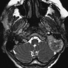
Figure 1
Figure 1
Magnetic resonance axial T2-weighted image of patient 1 with a giant lesion of the left temporal bone, with no obvious invasion of the parenchyma of the brain.
Figure 2
Figure 2
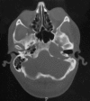
Computed tomography scan of patient 2 showing massive bone destruction of the skull base on the left side.
Figure 3
Figure 3
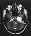
Magnetic resonance axial T2-weighted image of patient 2 with a hyperintense lesion of the left skull base.
Figure 4
Figure 4
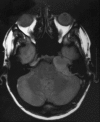
Magnetic resonance axial T1-weighted image of patient 2, showing a mass of the left skull base, isointense to gray matter.
Figure 5
Figure 5
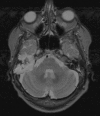
Magnetic resonance axial T2-weighted image of patient 3 showing a large mass in the temporal bone on the right side, hyperintense to gray matter.
Figure 6
Figure 6
Coronal computed tomography scan image of patient 3 showing massive bone destruction on the right side.
Figure 7
Figure 7
Axial computed tomography scan image of patient 4 showing the lesion on the right side, extending onto the sella and the sphenoidal sinus.
Figure 8
Figure 8
Axial computed tomography scan image of patient 5 revealing a giant lesion, covering almost one-third of the skull base on the left side.
Figure 9
Figure 9
Photograph taken of the skull base of patient 5 during autopsy. The extensive destruction of the skull base with the remains of the cholesteatoma is visible (left side).

References

    1. Kapur T R, Jayarmachandran S. Management of acquired cholesteatoma of the middle ear and the mastoid by combined approach tympanoplasty: a long-term view. Clin Otolaryngol Allied Sci. 1997;22:57–61. - PubMed
    1. Kinney S E. Intact canal wall tympanoplasty with mastoidectomy for cholesteatoma: long term follow-up. Laryngoscope. 1988;98:1190–1194. - PubMed
    1. Lau T, Tos M. Cholesteatoma in children. Recurrence related to observation period. Am J Otolaryngol. 1987;8:364–375. - PubMed
    1. Vartiainen E. Ten-years result of canal wall down mastoidectomy for acquired cholesteatoma. Auris Nasus Larynx. 2000;27:227–229. - PubMed
    1. Tos M, Lau T. Attic cholesteatoma. Recurrence rate related to observation time. Am J Otol. 1988;9:456–464. - PubMed

Publication types