

Solution structure of the cysteine-rich domain in Fn14, a member of the tumor necrosis factor receptor superfamily
- PMID: 19241374
- PMCID: PMC2760370
- DOI: 10.1002/pro.49
Solution structure of the cysteine-rich domain in Fn14, a member of the tumor necrosis factor receptor superfamily
Abstract
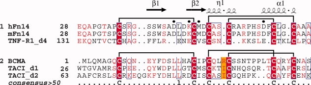
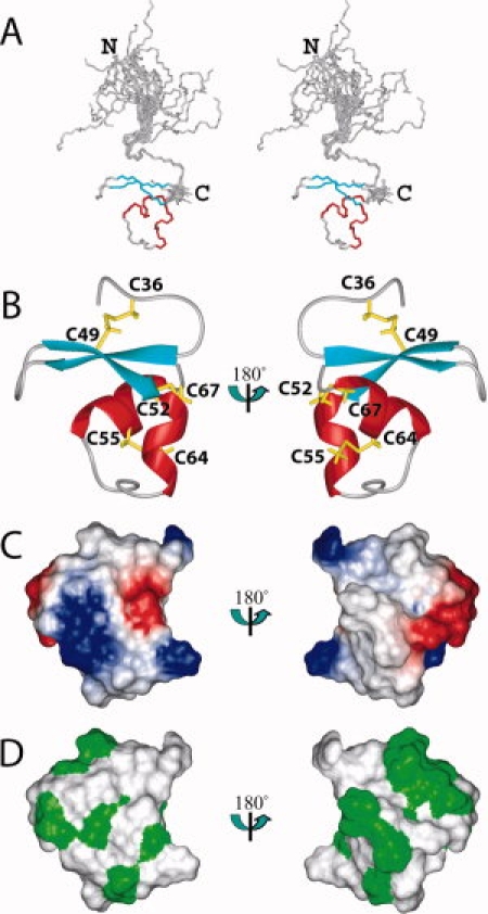
Fn14 is the smallest member of the tumor necrosis factor (TNF) receptor superfamily, and specifically binds to its ligand, TWEAK (TNF-like weak inducer of apoptosis), which is a member of the TNF superfamily. The receptor-ligand recognition between Fn14 and TWEAK induces a variety of cellular processes for tissue remodeling and is also involved in the pathogenesis of some human diseases, such as cancer, chronic autoimmune diseases, and acute ischaemic stroke. The extracellular ligand-binding region of Fn14 is composed of 53 amino acid residues and forms a single, cysteine-rich domain (CRD). In this study, we determined the solution structure of the Fn14 CRD (Glu28-Ala70) by heteronuclear NMR, with a (13)C-/(15)N-labeled sample. The tertiary structure of the CRD comprises a beta-sheet with two strands, followed by a 3(10) helix and a C-terminal alpha-helix, and is stabilized by three disulfide bonds connecting Cys36-Cys49, Cys52-Cys67, and Cys55-Cys64. Comparison of the disulfide bond connectivities and the tertiary structures with those of other CRDs revealed that the Fn14 CRD is similar to the fourth CRD of TNF receptor 1 (A1-C2 module type), but not to the CRD of B-cell maturation antigen and the second CRD of transmembrane activator and CAML (calcium modulator and cyclophilin ligand) interactor (A1-D2 module type). This is the first structural report about the A1-C2 type CRD that could bind to the known target.
Figures

Similar articles
-
TWEAK-independent Fn14 self-association and NF-κB activation is mediated by the C-terminal region of the Fn14 cytoplasmic domain.PLoS One. 2013 Jun 4;8(6):e65248. doi: 10.1371/journal.pone.0065248. Print 2013. PLoS One. 2013. PMID: 23750247 Free PMC article.
-
TWEAK binding to the Fn14 cysteine-rich domain depends on charged residues located in both the A1 and D2 modules.Biochem J. 2006 Jul 15;397(2):297-304. doi: 10.1042/BJ20051362. Biochem J. 2006. PMID: 16526941 Free PMC article.
-
BAFF/BLyS receptor 3 comprises a minimal TNF receptor-like module that encodes a highly focused ligand-binding site.Biochemistry. 2003 May 27;42(20):5977-83. doi: 10.1021/bi034017g. Biochemistry. 2003. PMID: 12755599
-
Role of TWEAK and Fn14 in tumor biology.Front Biosci. 2007 Jan 1;12:2761-71. doi: 10.2741/2270. Front Biosci. 2007. PMID: 17127278 Review.
-
TWEAK, a member of the TNF superfamily, is a multifunctional cytokine that binds the TweakR/Fn14 receptor.Cytokine Growth Factor Rev. 2003 Jun-Aug;14(3-4):241-9. doi: 10.1016/s1359-6101(03)00019-4. Cytokine Growth Factor Rev. 2003. PMID: 12787562 Review.
Cited by
-
TWEAK/Fn14 and Non-Canonical NF-kappaB Signaling in Kidney Disease.Front Immunol. 2013 Dec 10;4:447. doi: 10.3389/fimmu.2013.00447. Front Immunol. 2013. PMID: 24339827 Free PMC article. Review.
-
TWEAK and the progression of renal disease: clinical translation.Nephrol Dial Transplant. 2014 Feb;29 Suppl 1(Suppl 1):i54-i62. doi: 10.1093/ndt/gft342. Nephrol Dial Transplant. 2014. PMID: 24493870 Free PMC article. Review.
-
TWEAK-independent Fn14 self-association and NF-κB activation is mediated by the C-terminal region of the Fn14 cytoplasmic domain.PLoS One. 2013 Jun 4;8(6):e65248. doi: 10.1371/journal.pone.0065248. Print 2013. PLoS One. 2013. PMID: 23750247 Free PMC article.
-
The TWEAK/Fn14/CD163 axis-implications for metabolic disease.Rev Endocr Metab Disord. 2022 Jun;23(3):449-462. doi: 10.1007/s11154-021-09688-4. Epub 2021 Sep 20. Rev Endocr Metab Disord. 2022. PMID: 34542797 Free PMC article. Review.
-
Binding efficiency of protein-protein complexes.Biochemistry. 2012 Nov 13;51(45):9124-36. doi: 10.1021/bi301039t. Epub 2012 Nov 1. Biochemistry. 2012. PMID: 23088250 Free PMC article.
References
-
- Locksley RM, Killeen N, Lenardo MJ. The TNF and TNF receptor superfamilies: integrating mammalian biology. Cell. 2001;104:487–501. - PubMed
-
- Bodmer JL, Schneider P, Tschopp J. The molecular architecture of the TNF superfamily. Trends Biochem Sci. 2002;27:19–26. - PubMed
-
- Chicheportiche Y, Bourdon PR, Xu H, Hsu YM, Scott H, Hession C, Garcia I, Browning JL. TWEAK, a new secreted ligand in the tumor necrosis factor family that weakly induces apoptosis. J Biol Chem. 1997;272:32401–32410. - PubMed
-
- Marsters SA, Sheridan JP, Pitti RM, Brush J, Goddard A, Ashkenazi A. Identification of a ligand for the death-domain-containing receptor Apo3. Curr Biol. 1998;8:525–528. - PubMed
-
- Meighan-Mantha RL, Hsu DK, Guo Y, Brown SA, Feng SL, Peifley KA, Alberts GF, Copeland NG, Gilbert DJ, Jenkins NA, Richards CM, Winkles JA. The mitogen-inducible Fn14 gene encodes a type I transmembrane protein that modulates fibroblast adhesion and migration. J Biol Chem. 1999;274:33166–33176. - PubMed
Publication types
MeSH terms
Substances
Associated data
- Actions
LinkOut - more resources
Full Text Sources
Molecular Biology Databases
Research Materials
Miscellaneous
