. 2009 Mar;18(3):579-94.
doi: 10.1002/pro.64.

Protease inhibitors derived from elafin and SLPI and engineered to have enhanced specificity towards neutrophil serine proteases

Affiliations

Protease inhibitors derived from elafin and SLPI and engineered to have enhanced specificity towards neutrophil serine proteases

Marie-Louise Zani et al. Protein Sci. 2009 Mar.

Abstract

The secretory leukocyte protease inhibitor (SLPI), elafin, and its biologically active precursor trappin-2 are endogeneous low-molecular weight inhibitors of the chelonianin family that control the enzymatic activity of neutrophil serine proteases (NSPs) like elastase, proteinase 3, and cathepsin G. These inhibitors may be of therapeutic value, since unregulated NSP activities are linked to inflammatory lung diseases. However SLPI inhibits elastase and cathepsin G but not proteinase 3, while elafin targets elastase and proteinase 3 but not cathepsin G. We have used two strategies to design polyvalent inhibitors of NSPs that target all three NSPs and may be used in the aerosol-based treatment of inflammatory lung diseases. First, we fused the elafin domain with the second inhibitory domain of SLPI to produce recombinant chimeras that had the inhibitory properties of both parent molecules. Second, we generated the trappin-2 variant, trappin-2 A62L, in which the P1 residue Ala is replaced by Leu, as in the corresponding position in SLPI domain 2. The chimera inhibitors and trappin-2 A62L are tight-binding inhibitors of all three NSPs with subnanomolar K(i)s, similar to those of the parent molecules for their respective target proteases. We have also shown that these molecules inhibit the neutrophil membrane-bound forms of all three NSPs. The trappin-2 A62L and elafin-SLPI chimeras, like wild-type elafin and trappin-2, can be covalently cross-linked to fibronectin or elastin by a tissue transglutaminase, while retaining their polypotent inhibition of NSPs. Therefore, the inhibitors described herein have the appropriate properties to be further evaluated as therapeutic anti-inflammatory agents.


Figures

Figure 1
Figure 1
Sequence alignment of the regions surrounding inhibitory loops of elafin and the SLPI1 and SLPI2 domains. P3 to P3′ residues of the inhibitory loop are indicated. Residues that are conserved between at least two sequences are colored grey. The amino acids in elafin are numbered according to the trappin-2 sequence.
Figure 2
Figure 2
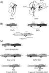
Diagram of the structures of engineered inhibitors. (A) Ribbon representation of the three-dimensional structure of elafin and SLPI extracted from the elafin-PPE complex coordinate file (PDB code: 1FLE) and SLPI-bovine chymotrypsin complex coordinate file (Dr. Bode) respectively. The four disulfide bonds in each WAP domain are shown as sticks. The P1 residue in each inhibitory loop is indicated by an asterisk. The figure was generated using PyMOL (http://www.pymol.org). (B) Structural organization of chelonianin inhibitors. Elafin is a 57-amino acid inhibitor (HNE and Pr3 inhibitor) derived from its active precursor trappin-2 (95 residues) by a proteolytic cleavage thought to involve mast cell tryptase (17). The N-terminal cementoïn domain of trappin-2 contains several repeated motifs rich in Gln and Lys residues that serve as transglutaminase substrates. The elafin domain is structurally homologous to both the SLPI domains (SLPI1 is N-terminal and SLPI2 is C-terminal). Only SLPI2 is believed to inhibit HNE and CatG, while SLPI1 inhibits trypsin. Also shown are the four disulfide bonds (bold lines) in each inhibitory (WAP) domain and the inhibitory loop of each WAP domain. (C) Structures of the inhibitors designed in this study. SLPI1(Elaf)-SLPI2 corresponds to SLPI in which the inhibitory loop of SLPI1 (amino acids 10 to 26) is replaced by the corresponding region of elafin (54–70). The Elaf-SLPI2 and SLPI2-Elaf chimeras combine the NSP-inhibiting properties of elafin (HNE and Pr3 inhibition) and SLPI2 (HNE and CatG inhibition). Trappin-2 A62L and trappin-2 R60Q/A62L/R69F are trappin-2 mutants in which residues P3, P1, and P7′ of the inhibitory loop are replaced by the corresponding residues in SLPI2.
Figure 3
Figure 3
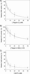
Dose dependent inhibition of membrane-bound NSPs. Curves showing the inhibition of HNE (A), Pr3 (B) and CatG (C) bound to the membrane of activated neutrophils by WT trappin-2, trappin-2 A62L and WT SLPI. Purified PMNs were activated with the calcium ionophore A23187 and then incubated for 30 min at 37°C with various concentrations of inhibitor. The number of cells was adjusted so that the concentration of each protease was 2 nM, as assessed by the hydrolysis of specific fluorogenic substrates. The residual protease activity was measured using specific fluorogenic substrates. Results are expressed as relative activity compared to the control experiment without added inhibitor and are the means ± SD of five separate experiments.
Figure 4
Figure 4
Sensitivities of membrane-bound NSPs to WT chelonianins and engineered inhibitors derived from elafin/trappin-2 or SLPI. Purified PMNs were activated with the calcium ionophore A23187 and then incubated for 30 min at 37°C with 100 nM of: (1) WT elafin, (2) WT trappin-2, (3) WT SLPI, (4) Elaf-SLPI2, (5) SLPI2-Elaf and (6) trappin-2 A62L variant. Residual enzyme activity of each membrane-bound protease was measured using specific fluorogenic substrates. Data are expressed as percentage of residual enzyme activity (rate of substrate hydrolysis in the presence of inhibitor to the rate of substrate hydrolysis without inhibitor) and are the means of three separate experiments. Like their soluble forms, membrane-bound CatG and Pr3 were not inhibited by WT elafin/trappin-2 or WT SLPI respectively. The engineered inhibitors also inhibited all three serine proteases bound at the surface of neutrophils, as they do the soluble proteases (Table I).
Figure 5
Figure 5
Inhibition of soluble NSPs by inhibitors cross-linked to fibronectin or elastin by transglutamination. Inhibitors were cross-linked to fibronectin or elastin in 96-well microplates essentially as described in (18). Inhibitors (10−6M) were incubated with tissue transglutaminase for 2 h at 37°C to form conjugated complexes with fibronectin (A,C) or elastin (B, D). The wells were washed thoroughly to remove unreacted products, and incubated with HNE (1 nM), Pr3 (2 nM), or CatG (2 nM) for 15 min at 37°C to allow protease-inhibitor complex formation. Residual enzyme activity was monitored using specific fluorogenic substrates. Representative inhibition curves (substrate hydrolysis expressed as fluorescence units vs time) are shown for the inhibition of HNE by Elaf-SLPI2 or trappin-2 A62L bound to fibronectin (A) or elastin (B). (C), (D), Graph show the residual enzyme activity for various protease-immobilized inhibitor pairs after cross-linking of the inhibitor to fibronectin (C) or elastin (D), calculated as the ratio of the rate of substrate hydrolysis in the presence of inhibitor to the rate of substrate hydrolysis without inhibitor (Control). Data are means ± SD for three separate experiments. The polyvalent inhibitors, Elaf-SLPI2 and trappin-2 A62L, inhibited all three NSPs when cross-linked to fibronectin or elastin, although Elaf-SLPI2 appeared to be less efficient against Pr3 than against HNE and CatG. As expected elafin and trappin-2 bound to fibronectin or elastin inhibited HNE and Pr3 but not CatG.
Figure 6
Figure 6
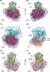
Electrostatic surface potentials of various protease-inhibitor complexes involving chelonianins. The atomic coordinates of elafin and SLPI extracted from the elafin-PPE and SLPI-chymotrypsin complexes were used to build theoretical complexes by superimposing elafin or SLPI and CatG or Pr3 onto the inhibitor or protease component of these X-ray complexes. The electrostatic potentials were calculated for the individual molecules of each complex using the Poisson-Boltzmann method implemented in DELPHI assuming a dielectric constant of two for the interior of the proteins and 80 for the exterior, an ionic strength of 0.15 M at pH 8.0 and a temperature of 300 K. The isopotential contours of positive or negative electrostatic potentials are displayed at +5 kTe−1 (blue for the protease, cyan for the inhibitor) and −5 kTe−1 (red for the protease, magenta for the inhibitor) respectively. Each complex is displayed with inhibitor at the top and protease at the bottom of each panel. CatG, with a net charge of +23, is entirely covered with a positive potential except for the active site entrance, which is negative, as in most serine proteases. Elafin may be prevented from interacting with CatG (A, B) because of positively charged residues: Arg60I of elafin (trappin-2 numbering) on one side of the complex (A) and Arg69I on the other side overlap with the positive regions of CatG in the reconstructed complex. For clarity, the Connolly surface of elafin is also shown and the CatG structure is shown as a solid ribbon. There are no unfavorable overlaps between SLPI (green ribbon) and CatG (white ribbon) in the reconstructed SLPI-CatG complex (C), despite both proteins having a high positive charge (SLPI net charge +12). Pr3 does not interact with SLPI, although it is almost neutral (net charge +1). The electrostatic contours of each component in the theoretical complex (D) reveal that two positive areas of Pr3, contributed by Arg36E/Arg65E and Lys99E/Arg168E that protrude towards SLPI and overlap with its positive surface. (E, F) the electrostatic contours for elafin and PPE in the X-ray structure of elafin-PPE complex show that complementary (favorable) contacts may help complex formation.

Similar articles

Cited by

References

    1. Shapiro SD. Proteinases in chronic obstructive pulmonary disease. Biochem Soc Trans. 2002;30:98–102. - PubMed
    1. Shapiro SD. Proteolysis in the lung. Eur Respir J Suppl. 2003;44:30s–32s. - PubMed
    1. Owen CA. Roles for proteinases in the pathogenesis of chronic obstructive pulmonary disease. Int J Chron Obstruct Pulmon Dis. 2008;3:253–268. - PMC - PubMed
    1. Voynow JA, Fischer BM, Zheng S. Proteases and cystic fibrosis. Int J Biochem Cell Biol. 2008;40:1238–1245. - PMC - PubMed
    1. Ganz T, Selsted ME, Szklarek D, Harwig SS, Daher K, Bainton DF, Lehrer RI. Defensins. Natural peptide antibiotics of human neutrophils. J Clin Invest. 1985;76:1427–1435. - PMC - PubMed

Publication types

MeSH terms

LinkOut - more resources