. 2009 Oct;30(10):3172-87.
doi: 10.1002/hbm.20739.

Probabilistic topography of human corpus callosum using cytoarchitectural parcellation and high angular resolution diffusion imaging tractography

Affiliations

Probabilistic topography of human corpus callosum using cytoarchitectural parcellation and high angular resolution diffusion imaging tractography

Yi-Ping Chao et al. Hum Brain Mapp. 2009 Oct.

Abstract

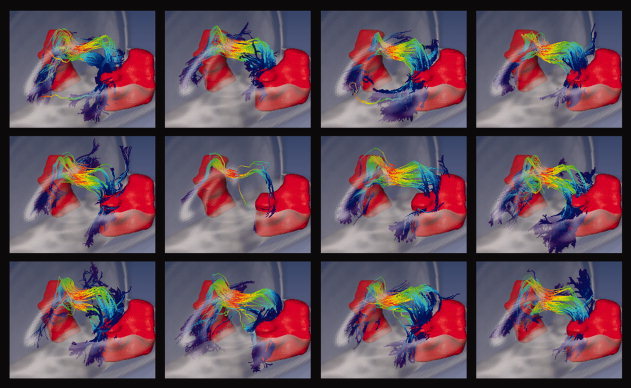
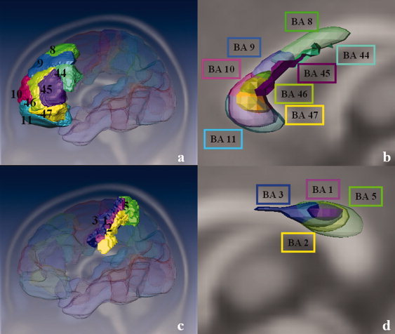
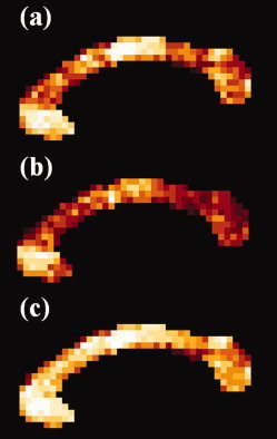
The function of the corpus callosum (CC) is to distribute perceptual, motor, cognitive, learned, and voluntary information between the two hemispheres of the brain. Accurate parcellation of the CC according to fiber composition and fiber connection is of upmost important. In this work, population-based probabilistic connection topographies of the CC, in the standard Montreal Neurological Institute (MNI) space, are estimated by incorporating anatomical cytoarchitectural parcellation with high angular resolution diffusion imaging (HARDI) tractography. First, callosal fibers are extracted using multiple fiber assignment by continuous tracking algorithm based on q-ball imaging (QBI), on 12 healthy and young subjects. Then, the fiber tracts are aligned in the standard MNI coordinate system based on a tract-based transformation scheme. Next, twenty-eight Brodmann's areas on the surface of cortical cortex are registered to the MNI space to parcellate the aligned callosal fibers. Finally, the population-based topological subdivisions of the midsagittal CC to each cortical target are then mapped. And the resulting subdivisions of the CC that connect to the frontal and somatosensory associated cortex are also showed. To our knowledge, it is the first topographic subdivisions of the CC done using HARDI tractography and cytoarchitectonic information. In conclusion, this sophisticated topography of the CC may serve as a landmark to further understand the correlations between the CC, brain intercommunication, and functional cytoarchitectures.


Figures

Figure 1
Figure 1
Flowchart of the tract‐based transformation. Fiber tracts reconstructed from individual native space are transformed to the standard MNI coordinate system using the spatial transformation function, which was derived from registration between the resliced TIWI and the ICBM‐152 T1WI. This allowed for group analysis of the CC topography derived from cytoarchitectural parcellation. The tracts are color‐coded according to the distance between seed point and target voxel. [Color figure can be viewed in the online issue, which is available at www.interscience.wiley.com.]
Figure 2
Figure 2
Callosal fibers of a single subject. (a) Callosal fibers extracted by MFACT with QBI overlaid onto the individual's anatomical image (T1WI). (b) The spatial normalized callosal fibers superimposed on the ICBM‐152 T1 image. The red rectangles in (a,b) show the homologous spatial relationship between individual native space and the MNI coordinate. The neural connections were well preserved after the tract‐based transformation. (c) An example of the neural connections between the CC and BA, showing the transformed callosal fibers projecting into the primary motor cortex (BA 4). [Color figure can be viewed in the online issue, which is available at www.interscience.wiley.com.]
Figure 3
Figure 3
Three‐dimensional (3D) callosal connections projecting to lateral and inferior BAs. Fiber pathways were clustered according to their projecting targets, temporal lobes (BAs 20–22, 37, 38, and 41/42) and the lateral regions (BAs 43 and 39) of human brain respectively in a single subject. Integrating QBI and MFACT algorithm, fiber tracts between the CC and temporal as well as lateral regions can be identified. [Color figure can be viewed in the online issue, which is available at www.interscience.wiley.com.]
Figure 4
Figure 4
Callosal fibers connected to BA 37 (occipito‐temporal area) were overlaid on the ICBM‐152 T1 image in the MNI coordinate. The tracts are consistent between all subjects (n = 12). [Color figure can be viewed in the online issue, which is available at www.interscience.wiley.com.]
Figure 5
Figure 5
Probabilistic topography of the midsagittal CC. Twenty‐eight BAs located at the surface of the cerebrum were considered as the terminal regions for cytoarchitecture parcellation. The CC was partitioned into subdivisions based on their commissural connections to the corresponding BAs, and then a population‐based probability topography of CC was constructed from 12 subjects. The color scale shown at the left bottom represents the population probability of a given voxel within the CC projecting to a particular BA. [Color figure can be viewed in the online issue, which is available at www.interscience.wiley.com.]
Figure 6
Figure 6
3D rendering of the sophisticated subdivisions in the CC based on its connections to the frontal and somatosensory associated BAs. Sagittal view of 3D reconstruction of major callosal distributions connected to the selected frontal BAs (BAs 8–11 and 44–47) (a) and somatosensory associated cortex (BAs 1–3 and 5) (c) were shown by thresholding those voxels in which greater than 50 and 20% of the population probability, respectively (b and d). Arbitary thresholds were selected to highlight the major callosal distributions based on their complexities and consistencies of tracking pathways. [Color figure can be viewed in the online issue, which is available at www.interscience.wiley.com.]
Figure 7
Figure 7
Hard segmentation of the midsagittal corpus callosum. BAs were reassigned to seven cortical targets, frontal cortex (green), premotor and supplementary motor areas (light blue), primary motor cortex (dark blue), primary sensory cortex (red), parietal lobe (orange), occipital lobe (yellow), and temporal lobe (violet) (a). The global topography of each subject was constructed using the hard segmentation method (b). In comparison with Witelson's scheme (top), our proposed scheme (middle), and Hofer's scheme (bottom) of the CC classification were shown (c). A geometric baseline was defined by connecting the most anterior (left) and posterior (right) points of the CC. According to geometrical baselines defined by Witelson [1989], five vertical partitions of the CC (top) were defined as anterior third (prefrontal, premotor and supplementary motor), anterior midbody (primary motor), posterior midbody (somaesthetic, posterior parietal), isthmus (posterior parietal, superior temporal), and splenium (occipital, inferior temporal). Similar to Witelson, five vertical partitions of the CC (bottom) defined by Hofer and Frahm [2006] were first sixth (prefrontal), the rest of the anterior half of the CC (premotor and supplementary motor), posterior half minus the posterior third (motor), posterior one‐third minus posterior one‐fourth (sensory), and posterior one‐fourth (parietal, temporal and occipital). Our five vertical partitions of the CC (middle) were defined as anterior one‐third (frontal), middle one‐third (premotor and supplementary motor), posterior one‐third minus the posterior one‐fourth (motor), posterior one‐fourth minus posterior one‐sixth (sensory), and posterior one‐sixth (parietal, temporal and occipital). [Color figure can be viewed in the online issue, which is available at www.interscience.wiley.com.]
Figure 8
Figure 8
The correspondence map of the primary (a) and the secondary (b) dominant BA label of each CC voxel between 12 subjects. The color of each voxel was identical to that of Figure 5. It was obvious that either of the primary or the secondary dominant cytoarchitectural label was not sufficient to show reliability of dominant CC connection map. However, integrating both the primary and secondary most dominant cytoarchitectonic labels resulted in a consistent correspondence map among all individuals (c). [Color figure can be viewed in the online issue, which is available at www.interscience.wiley.com.]

Similar articles

Cited by

References

    1. Abe O,Masutani Y,Aoki S,Yamasue H,Yamada H,Kasai K,Mori H,Hayashi N,Masumoto T,Ohtomo K ( 2004): Topography of the human corpus callosum using diffusion tensor tractography. J Comput Assist Tomogr 28: 533–539. - PubMed
    1. Alexander DC ( 2005): Multiple‐fiber reconstruction algorithms for diffusion MRI. Ann N Y Acad Sci 1064: 113–133. - PubMed
    1. Alexander DC,Pierpaoli C,Basser PJ,Gee JC ( 2001): Spatial transformations of diffusion tensor magnetic resonance images. IEEE Trans Med Imaging 20: 1131–1139. - PubMed
    1. Alexander AL,Lee JE,Lazar M,Boudos R,DuBray MB,Oakes TR,Miller JN,Lu J,Jeong EK,McMahon WM,Bigler ED,Lainhart JE ( 2007): Diffusion tensor imaging of the corpus callosum in Autism. Neuroimage 34: 61–73. - PubMed
    1. Amunts K,Malikovic A,Mohlberg H,Schormann T,Zilles K ( 2000): Brodmann's areas 17 and 18 brought into stereotaxic space—Where and how variable? Neuroimage 11: 66–84. - PubMed