. 2009;4(2):e4612.
doi: 10.1371/journal.pone.0004612. Epub 2009 Feb 26.

Dissection of kinesin's processivity

Affiliations

Dissection of kinesin's processivity

Sarah Adio et al. PLoS One. 2009.

Abstract

The protein family of kinesins contains processive motor proteins that move stepwise along microtubules. This mechanism requires the precise coupling of the catalytic steps in the two heads, and their precise mechanical coordination. Here we show that these functionalities can be uncoupled in chimera of processive and non-processive kinesins. A chimera with the motor domain of Kinesin-1 and the dimerization domain of a non-processive Kinesin-3 motor behaves qualitatively as conventional kinesin and moves processively in TIRF and bead motility assays, suggesting that spatial proximity of two Kinein-1 motor domains is sufficient for processive behavior. In the reverse chimera, the non-processive motor domains are unable to step along microtubules, despite the presence of the Kinesin-1 neck coiled coil. Still, ATP-binding to one head of these chimera induces ADP-release from the partner head, a characteristic feature of alternating site catalysis. These results show that processive movement of kinesin dimers requires elements in the motor head that respond to ADP-release and induce stepping, in addition to a proper spacing of the motor heads via the neck coiled coil.


Conflict of interest statement

Competing Interests: The authors have declared that no competing interests exist.

Figures

Figure 1
Figure 1. Design of chimeric kinesin constructs.
The upper part of the figure shows the domain organization and the fusion points of chimeric kinesin motors. Below, the neck regions are shown in detail. The coiled coil assignment for the kinesin-1 members NcKin and HsKIF5B (ubiquitous conventional kinesin) is taken from the crystal structure 3KIN, the NcKin3 coiled coil is predicted by computer algorithms and experimental data . Positively charged residues are blue, negatively charged red.
Figure 2
Figure 2. Single-molecule properties of the Head1/Neck3 chimera.
(A) Kymographs show displacements of a single fluorophore-labeled Head1/Neck3 motor along the microtubule. (B) Wildtype NcKin was used as a control. A single motor moves continuously on a microtubule with a velocity essentially identical to the average velocity under multiple motor conditions. Motors were observed for 5 sec with an integration time of 200 ms at an ATP concentration of 20 µM. (C) Record of a microscopic latex bead captured by an optical trap and attached to a single Head1/Neck3 protein. Upon binding to surface immobilized microtubules the motor moves stepwise along the filament until it detaches at a stall force of 3.1 pN. Near stall resolution of 8 nm steps indicate hand-over-hand motility. (D) Head1/Neck3 velocities under load. Average velocities v (mean±s.e.m.) of NcKin wild type motor (closed circles and additional data points) and Head1/Neck3 chimera (open circles) are plotted versus applied load. Measurements were performed under constant force using a force feedback-controlled optical trap. The average velocities of both motors drop with increasing external force. Data were fitted (solid line) by Bell's equation assuming a kinetic model with one force-independent rate-limiting transition, and one force-dependent rate. The transition state position of approximately 8 nm suggests that the step is dominated by diffusive search. (E) Head1/Neck3 runlength under load. Average runlengths (mean±SE) of NcKin wild type motor (closed circles, data from , model according to [47]) chimera (open circles) are plotted versus applied load. At all forces tested the chimera has a reduced runlength compared to wild type NcKin. Imperfect head-head coordination is most likely the reason for a higher detachment probability and the decrease in processivity.
Figure 3
Figure 3. kbi ratio of non-processive Head3/Neck1.
(A) The steady state ATPase rate of the Head3/Neck1 motor was measured at variable microtubule concentrations. Based on the concentrations of polypeptide chains the kcat was 23.2 s−1 and the microtubule concentration for half maximal activation (K0.5,MT) was 0.2 µM, the apparent binding rate between Head3/Neck1 and microtubules (kbi(ATP) = kcat/K0.5,MT) was 116.0 µM−1·s−1 (Table 1). (B) To determine the ADP release rate mant-ADP loaded Head3/Neck1 was mixed with microtubules in a stopped-flow apparatus. The left inset shows an example of the fluorescence decay at 1.2 µM microtubules. The reaction was fitted with a double-exponential function (grey line). Each data point is an average of at least five individual stopped-flow traces. Rates were plotted against the microtubule concentration. The hyperbolic fit of the data revealed a maximal ADP release rate of kmax = 52.5±4.5 s−1 with a K0.5, MT = 1.72±0.43 µM for the fast rate, and kmax = 0.65±0.02 s−1/K0.5, MT = 0.53±0.08 µM for the slow phase (right inset). The comparison of the bimolecular rate of the fast rate in this assay (kmax/K0.5,MT = 30.5 µM−1 s−1) with the apparent rate in steady state reveals a low biochemical processivity index of 3–4 ATPs hydrolyzed per microtubule encounter by the Head3/Neck1 chimera.
Figure 4
Figure 4. Microtubule activated ADP release from the Head3/Neck1 chimera.
Panel A shows the normalized time courses of mantADP release from Head3/Neck1-mantADP complex after addition of various amount of microtubules (final concentration c = 0.25 µM closed circles, c = 0.5 µM grey circles and c = 1.0 µM open circles) and 1 mM ATP. Panel B summarizes the fluorescence amplitudes after addition of microtubules (closed circles) and ATP (open circles).
Figure 5
Figure 5. Dissociation of the Head3/Neck1 microtubule complex.
The dissociation of the pre-formed Head3/Neck1 microtubule complex was induced by ATP in a stopped-flow apparatus (inset panel A) and followed by the change of the light scattering signal. (A) The graph shows a representative average from 5 traces. The grey curve is a mono-exponential fit to the data that was used to derive kobs. (B) Graph B shows the hyperbolic dependence of kobs on the ATP concentration, with a kmax of 0.47 s−1 and a K1/2 of 105.0 µM ATP.
Figure 6
Figure 6. Kinetic models of constructs of this study.
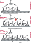
The figure summarizes the different mechanisms of processive and non-processive kinesins. (A) The non-processive Kinesin-3 reference construct NcKin3 binds to microtubules and detaches after one hydrolysis cycle. (B) The Head3/Neck1 chimera is unable to detach from the filament after one catalytic cycle (intermediate 5). At this state, the kinetic pathway branches and part of the enzymes cycle through ATP hydrolysis, part of them dwell in a long-lived microtubule-bound state (6′) before detachment. (C) NcKin and Head1/Neck3 mutant are processive enzymes that proceed after intermediate 5 via a double-head bound intermediate to the initial state where they are able to repeat the catalytic cycle.

Similar articles

Cited by

References

    1. Vale RD, Reese TS, Sheetz MP. Identification of a novel force-generating protein, kinesin, involved in microtubule-based motility. Cell. 1985;42:39–50. - PMC - PubMed
    1. Howard J, Hudspeth AJ, Vale R. Movement of microtubules by single kinesin molecules. Nature. 1989;342:154–158. - PubMed
    1. Hackney D. Evidence for alternating head catalysis by kinesin during microtubule-stimulated ATP hydrolysis. PNAS. 1994;91:6865–6869. - PMC - PubMed
    1. Crevel IM, Nyitrai M, Alonso MC, Weiss S, Geeves MA, et al. What kinesin does at roadblocks: the coordination mechanism for molecular walking. Embo J. 2004;23:23–32. - PMC - PubMed
    1. Cross RA. The kinetic mechanism of kinesin. Trends Biochem Sci. 2004;29:301–309. - PubMed

Publication types