. 2009;4(2):e4424.
doi: 10.1371/journal.pone.0004424. Epub 2009 Feb 10.

Depletion of kinesin 5B affects lysosomal distribution and stability and induces peri-nuclear accumulation of autophagosomes in cancer cells

Affiliations

Depletion of kinesin 5B affects lysosomal distribution and stability and induces peri-nuclear accumulation of autophagosomes in cancer cells

Carla M P Cardoso et al. PLoS One. 2009.

Abstract

Background: Enhanced lysosomal trafficking is associated with metastatic cancer. In an attempt to discover cancer relevant lysosomal motor proteins, we compared the lysosomal proteomes from parental MCF-7 breast cancer cells with those from highly invasive MCF-7 cells that express an active form of the ErbB2 (DeltaN-ErbB2).

Methodology/principal findings: Mass spectrometry analysis identified kinesin heavy chain protein KIF5B as the only microtubule motor associated with the lysosomes in MCF-7 cells, and ectopic DeltaN-ErbB2 enhanced its lysosomal association. KIF5B associated with lysosomes also in HeLa cervix carcinoma cells as analyzed by subcellular fractionation. The depletion of KIF5B triggered peripheral aggregations of lysosomes followed by lysosomal destabilization, and cell death in HeLa cells. Lysosomal exocytosis in response to plasma membrane damage as well as fluid phase endocytosis functioned, however, normally in these cells. Both HeLa and MCF-7 cells appeared to express similar levels of the KIF5B isoform but the death phenotype was weaker in KIF5B-depleted MCF-7 cells. Surprisingly, KIF5B depletion inhibited the rapamycin-induced accumulation of autophagosomes in MCF-7 cells. In KIF5B-depleted cells the autophagosomes formed and accumulated in the close proximity to the Golgi apparatus, whereas in the control cells they appeared uniformly distributed in the cytoplasm.

Conclusions/significance: Our data identify KIF5B as a cancer relevant lysosomal motor protein with additional functions in autophagosome formation.


Conflict of interest statement

Competing Interests: The authors have declared that no competing interests exist.

Figures

Figure 1
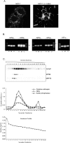
Figure 1. KIF5B is highly expressed in cancer cells and associates with lysosome containing fractions.
(A) Immunostaining with lysosome specific LAMP-1 antibody in ΔN-ErbB2 and control cells. (B) RT-PCR analysis showing the mRNA expression levels of various N-kinesins including KIF5A (349 bp), KIF5B (337 bp), KIF5C (320 bp), KIF3A (393 bp) and KIF1A (364 bp) in H: HeLa, M: MCF-7 and U: U2OS cells. KIF amplification bands are indicated with arrows. The house-keeping gene PBDG (257 bp) was used as internal control. (C) Light membrane fractions of HeLa cells were separated by iodixanol gradient ultracentrifugation and the protein expression levels of KIF5B, LAMP-2 (lysosomes) and GRP75 (mitochondria) were visualized by immunoblotting. The enzymatic activity levels of Cathepsin B/L, acidic phosphatase and NAG was measured in all fractions and served as lysosomal markers; the linearity of the iodixanol gradient profile was determined by measuring OD at 244 nm (lower graph).
Figure 2
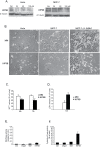
Figure 2. Depletion of KIF5B induces moderate cytoxicity in MCF-7 cells and massive cell death in HeLa cells.
(A) Protein levels of KIF5B, 48 hours after depletion with varying siRNA concentrations (KIF5B-1 siRNA) for HeLa and MCF-7; β-tubulin served as internal control. Oligof: control cells treated with oligofectamine alone. (B) Representative phase contrast pictures of HeLa, MCF-7 and MCF-7-ΔN-ErbB2 cells, 72 h after treatment with indicated siRNAs. (C) HeLa cells were depleted for KIF5B or treated with control MM siRNA and metabolic activity determined by a MTT assay and death estimated by LDH release assay (D). MTT and LDH values is presented as percentage of untreated cells. (E) Caspase-3 like activity and (F) cytosolic cathepsin activity in HeLa cells after KIF5B depletion (72 hours) measured by DEVDase and zFRase enzyme assays respectively. Values represent means of triplicate measurements ± SD. All experiments were repeated three times with essentially the same results.
Figure 3
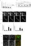
Figure 3. KIF5B depletion induces pericellular aggregation of lysosomes in HeLa cells but has no impact on exocytosis activity.
(A) Representative confocal pictures of either HeLa cells or HeLa stably expressing LIMP-1-EGFP transfected with KIF5B or MM siRNA, and stained with indicated antibodies. (B) HeLa cells seeded on coverslips (80% confluency) were membrane wounded with a scalpel and immediately after stained for surface LAMP-1; cells were subsequently fixated and stained for KIF5B. (C) HeLa cells transfected with indicated siRNAs were (after 48 h) stimulated to exocytose with 10 μM ionomycin. Extracellular secretion of lysosomal cathepsins was measured by a zFR-AMC enzyme assay and values (means of triplicate measurements ± SD) were expressed as percent of total cellular LDH content. (D) Quantification of surface LAMP-1 in electroporated HeLa cells by flow cytometry. Red and green indicates cells in two different gates. The percentage of cells in the red gate was used to estimate the amount of surface-exposed LAMP-1 +/− electroporation. FL1-H: fluorescence intensity. FSC-H: forward side scatter.
Figure 4
Figure 4. KIF5B depletion suppresses autophagy and induces nuclear accumulation of autophagosomes.
(A) MCF-7-LC3-eGFP cells treated with oligofectamine or transfected with indicated siRNAs were left untreated or stimulated with rapamycin for 24 h. The percentage of cells with LC3-eGFP localized to ≥five cytosolic granular structures was estimated by counting a minimum of 100 cells/sample. Values represent means of three independent experiments ± SD. (B) Immunoblots showing the protein levels after depletion (72 h) with indicated siRNAs or treatment with 1 μM rapamycin for 12 h. p70-s6K: phosphorylated form of p70 S6 kinase. eGFP-LC3: eGFP antibody specific for eGFP (fused to LC3) (C) Representative phase contrast pictures adapted from time lapse movies of MCF-7-LC3-eGFP cells treated with Concanamycin A (6 nM) to induce autophagy and LC3-eGFP translocation to autophagosomes, 72 h after transfection with indicated siRNAs. (D and E) MCF-7-LC3-eGFP cells transfected with indicated siRNAs were (after 72 h) imaged by time lapse microscopy during treatment with either 6 nM concanamycin A (D) or 4 μM rapamycin (E). The percentage of cells displaying nuclear accumulation of LC3-eGFP autophagosomes were scored after 45 min incubation. (F) Immuno staining of trans-Golgi (Golgin-97 Ab) in MCF-7-LC3-eGFP cells depleted for KIF5B and incubated for 45 min with concanamycin A. Values represent means of 3–4 independent experiments. P-values: MM/KIF5B. *: p<0,05; **: p<0,01; ***: p<0,001 (student's T-test).

Similar articles

Cited by

References

    1. Eskelinen EL, Tanaka Y, Saftig P. At the acidic edge: emerging functions for lysosomal membrane proteins. Trends Cell Biol. 2003;13:137–145. - PubMed
    1. Gieselmann V. Lysosomal storage diseases. Biochim Biophys Acta. 1995;1270:103–136. - PubMed
    1. Gocheva V, Joyce JA. Cysteine cathepsins and the cutting edge of cancer invasion. Cell Cycle. 2007;6:60–64. - PubMed
    1. Jedeszko C, Sloane BF. Cysteine cathepsins in human cancer. Biol Chem. 2004;385:1017–1027. - PubMed
    1. Fehrenbacher N, Gyrd-Hansen M, Poulsen B, Felbor U, Kallunki T, et al. Sensitization to the lysosomal cell death pathway upon immortalization and transformation. Cancer Res. 2004;64:5301–5310. - PubMed

Publication types