. 2009 Feb;5(2):e1000291.
doi: 10.1371/journal.ppat.1000291. Epub 2009 Feb 6.

HCV induces oxidative and ER stress, and sensitizes infected cells to apoptosis in SCID/Alb-uPA mice

Affiliations

HCV induces oxidative and ER stress, and sensitizes infected cells to apoptosis in SCID/Alb-uPA mice

Michael A Joyce et al. PLoS Pathog. 2009 Feb.

Abstract

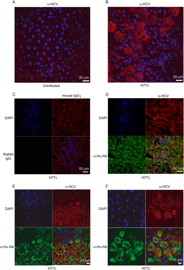
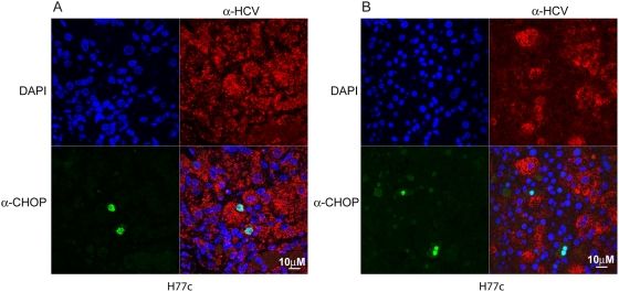
Hepatitis C virus (HCV) is a blood-borne pathogen and a major cause of liver disease worldwide. Gene expression profiling was used to characterize the transcriptional response to HCV H77c infection. Evidence is presented for activation of innate antiviral signaling pathways as well as induction of lipid metabolism genes, which may contribute to oxidative stress. We also found that infection of chimeric SCID/Alb-uPA mice by HCV led to signs of hepatocyte damage and apoptosis, which in patients plays a role in activation of stellate cells, recruitment of macrophages, and the subsequent development of fibrosis. Infection of chimeric mice with HCV H77c also led an inflammatory response characterized by infiltration of monocytes and macrophages. There was increased apoptosis in HCV-infected human hepatocytes in H77c-infected mice but not in mice inoculated with a replication incompetent H77c mutant. Moreover, TUNEL reactivity was restricted to HCV-infected hepatocytes, but an increase in FAS expression was not. To gain insight into the factors contributing specific apoptosis of HCV infected cells, immunohistological and confocal microscopy using antibodies for key apoptotic mediators was done. We found that the ER chaperone BiP/GRP78 was increased in HCV-infected cells as was activated BAX, but the activator of ER stress-mediated apoptosis CHOP was not. We found that overall levels of NF-kappaB and BCL-xL were increased by infection; however, within an infected liver, comparison of infected cells to uninfected cells indicated both NF-kappaB and BCL-xL were decreased in HCV-infected cells. We conclude that HCV contributes to hepatocyte damage and apoptosis by inducing stress and pro-apoptotic BAX while preventing the induction of anti-apoptotic NF-kappaB and BCL-xL, thus sensitizing hepatocytes to apoptosis.


Conflict of interest statement

The authors have declared that no competing interests exist.

Figures

Figure 1
Figure 1. Confocal microscopy of uninfected and HCV H77c infected mouse livers stained with anti-HCV and anti-human albumin.
Confocal microscopy was performed on either uninfected (A) or H77c infected (B–F) liver sections. Sections were either singly stained (A, B) using mouse monoclonal antibodies against HCV or, to differentiate between the human and mouse hepatocytes, rabbit anti human albumin antibodies in addition (D–F). Isotype controls (C) were also used to stain a serial section to panel D (See also figure S1 A, B). The nuclei were stained with DAPI (blue), and mouse antibodies were visualized using secondary goat anti-mouse poly-HRP antibodies followed by tyramide-TMR (red). The rabbit antibodies were visualized using secondary goat anti-rabbit-alexa 488 (green) antibodies. Junctions between human and mouse cells is shown in two magnifications in panels E and F.
Figure 2
Figure 2. In situ hybridization with human specific Alu DNA oligonucleotides.
In situ hybridization using human Alu DNA probes, with the nuclei were counter stained using methyl green, showing a lobular region (A), and portal triad (B). Magnification ×400.
Figure 3
Figure 3. Global gene expression profiles of HCV (H77c 1a)-infected mice.
Two-dimensional hierarchical clustering was performed using Resolver System software with an agglomerative algorithm, complete link heuristic criteria, and Euclidian correlation metric. Each column represents gene expression data from an individual experiment (either individual HCV-infected mouse or individual liver sample) and the cluster represents genes that showed a >2-fold change (P value<0.05) in at least 1 experiment. Genes shown in red were up-regulated; genes shown in green down-regulated and genes in black indicate no change in expression in HCV-infected tissue relative to a pool (3 individual animals) of donor-matched uninfected tissue. Animal 990 was infected with H77c (+) serum. Animal 975 received 100 µg of H77c RNA by intrahepatic injection while animal 986 received 100 µg of H77c RNA containing the GDD-AAA mutation in the NS5B coding region.
Figure 4
Figure 4. Expression of IFN-inducible and lipid metabolism genes in human liver tissue from HCV-infected mice.
The gene sets were IFN-inducible (A), or involved in lipid metabolism (B). Two-dimensional hierarchical clustering was performed using Resolver System software with an agglomerative algorithm, complete link heuristic criteria, and Euclidean correlation metric. Each column represents gene expression data from an individual experiment (either individual HCV-infected mouse or individual liver sample). Genes were selected as at least 2-fold regulated (P value<0.05) in at least 1 experiment. Genes shown in red are up-regulated and genes shown in green are down-regulated in HCV-infected tissue relative to donor-matched uninfected tissue, while black indicates no change in gene expression. Animal 990 was infected with H77c (+) serum. Animal 975 H77c RNA by intrahepatic injection while animal 986 received H77c RNA containing the GDD-AAA mutation in the NS5B coding region.
Figure 5
Figure 5. Immunohistochemical analysis of FAS expression and TUNEL reactivity.
Liver sections from PBS injected (A and D), replication deficient RNA injected (B and E) and HCV H77c infected (C and F) donor matched chimeric mice were stained using rabbit anti-FAS (A–C), developed using the Vecastain ABC kit and counterstained using haematoxylin. Isotype controls were negative and are shown in Figure S1C–D. TUNEL (D–F) was performed using the Apoptag Plus Peroxidase In Situ Apoptosis Detection kit and the nuclei were counterstained with methyl green. Magnification ×100. Arrows indicate TUNEL reactive nuclei. In panel F, arrows indicate only some of the TUNEL reactive nuclei.
Figure 6
Figure 6. Confocal microscopic analysis of HCV antigen expression and either FAS expression or TUNEL reactivity.
Staining for FAS (A) or TUNEL reactivity (B) was performed as described in Materials and Methods, using primary mouse anti-HCV and either rabbit anti-human FAS (A), or for TUNEL, FITC labeled dUTP and terminal deoxynucleotidyl transferase (B). The secondary antibodies were goat anti mouse poly-HRP, and for FAS staining, goat anti rabbit Alexa-488 (green). HRP was developed using the TSA plus fluorescence system with tyramide-TMR (red). Nuclei were stained using DAPI (blue). Arrows indicate TUNEL reactive nuclei.
Figure 7
Figure 7. Immunohistochemical and confocal microscopic analysis of BiP/Grp78 expression.
BiP expression was examined by either immunohistochemistry (A–C) or confocal microscopy (D–E). Liver sections from PBS injected (A and D), replication deficient RNA injected (B) and HCV H77c infected (C and E) donor matched chimeric mice were stained using goat anti-BiP/Grp78 alone (A–C) and developed using the Vecastain ABC kit. Magnification ×100. Isotype controls are shown in Figure S1 E–F and are negative. For confocal microscopy (D and E), primary antibodies were goat anti-BiP/Grp78 with mouse anti-HCV, and the secondary antibodies were donkey anti-goat alexa 488 (green), and donkey anti-mouse biotin followed by avidin-HRP (Vector laboratories). The peroxidase was developed as before using tyramide-TMR (red). Nuclei were stained using DAPI (blue).
Figure 8
Figure 8. Immunohistochemical and confocal microscopic analysis of BAX expression.
BAX expression was examined by either immunohistochemistry (A–D) or confocal microscopy (E–F). Liver sections from PBS injected (A and E), replication deficient RNA injected (B) and HCV H77c infected (C, D and F) donor matched chimeric mice were stained using rabbit anti-BAX alone (A–D) and developed using the Vecastain ABC kit. Magnification ×100 (A–C). A magnification ×400 view is shown in D with red arrows indicating cells diffuse BAX, and black arrows indicating cells with punctate BAX. Isotype controls are shown in Figure S3 and are negative. For confocal microscopy, primary antibodies were rabbit anti-BAX with mouse anti-HCV, and the secondary antibodies were goat anti-rabbit alexa 488 (green), with goat anti-mouse poly-HRP, developed using tyramide-TMR (red). Nuclei were stained using DAPI (blue). Comparison of a field from an area of liver containing only mouse cells with an HCV infected human area is shown in Figure S3.
Figure 9
Figure 9. Confocal microscopy of HCV and CHOP/GADD153 in predominately infected or uninfected areas of HCV infected mice.
Panel (A) shows an area from an HCV infected liver that contains mostly infected cells. Panel (B) shows an area from an HCV infected liver that contains mostly uninfected cells. Liver sections from HCV H77c infected mice were stained using rabbit anti-CHOP and mouse anti-HCV antibodies, and the secondary antibodies were goat anti rabbit alexa 488 (green) and goat anti mouse poly-HRP, which was developed using tyramide-TMR (red). Nuclei were stained using DAPI (blue).
Figure 10
Figure 10. Confocal microscopy of HCV and either NF-κB p65 or BCL-xL.
Liver sections from PBS injected (A and C) and HCV H77c infected (B and D) chimeric donor matched mice were stained using anti-NF-κB p65 (A and B) or BCL-xL (C and D) antibodies. Panels E and F are from non-donor matched infected mice stained with anti-NF-κB p65 (E) or BCL-xL (F) Primary antibodies were rabbit anti-NF-κB or rabbit anti BCL-xL with mouse anti-HCV, and the secondary antibodies were goat anti-rabbit alexa 488 (green), with goat anti-mouse poly-HRP developed using tyramide-TMR (red). Nuclei were stained using DAPI (blue). Red arrows indicate infected cells and white arrows indicate uninfected cells. The right hand panels are quantitation of p65 and BCL-xL levels in uninfected and HCV infected cells in HCV infected livers. For p65, 6 fields (61 infected and 59 uninfected cells) were quantified using Metamorph software. For BCL-xL, 4 fields (44 infected and 43 uninfected cells) were quantified. To compare cells from several fields, the average of the uninfected cells in a single field was arbitrarily set to 1 and the infected cells in that field were scaled appropriately. Isotype controls and comparison of a field from an area of liver containing only mouse cells with an HCV infected human area are shown in Figure S4.
Figure 11
Figure 11. HCV sensitizes infected hepatocytes to apoptosis.
The pathways that link oxidative and ER stress with apoptosis are shown, as is a potential interaction between the host cell response and apoptosis. In hepatocytes the levels of caspase 8 are low and induction of apoptosis requires the mitochondrial amplification loop, the convergence of the stress pathways at the mitochondria and low levels of NF-κB and BCL-xL sensitize hepatocytes to apoptosis. Proteins that we have shown are elevated in HCV infected cells are shown in red, those that do not change are shown in yellow and those that are decreased are shown in green.

Similar articles

Cited by

References

    1. Alter MJ, Margolis HS, Krawczynski K, Judson FN, Mares A, et al. The natural history of community-acquired hepatitis C in the United States. The Sentinel Counties Chronic non-A, non-B Hepatitis Study Team. N Engl J Med. 1992;327:1899–1905. - PubMed
    1. Roberts MS, Angus DC, Bryce CL, Valenta Z, Weissfeld L. Survival after liver transplantation in the United States: a disease-specific analysis of the UNOS database. Liver Transpl. 2004;10:886–897. - PubMed
    1. Timm J, Roggendorf M. Sequence diversity of hepatitis C virus: implications for immune control and therapy. World J Gastroenterol. 2007;13:4808–4817. - PMC - PubMed
    1. Bigger CB, Guerra B, Brasky KM, Hubbard G, Beard MR, et al. Intrahepatic gene expression during chronic hepatitis C virus infection in chimpanzees. J Virol. 2004;78:13779–13792. - PMC - PubMed
    1. Smith MW, Yue ZN, Korth MJ, Do HA, Boix L, et al. Hepatitis C virus and liver disease: global transcriptional profiling and identification of potential markers. Hepatology. 2003;38:1458–1467. - PubMed

Publication types

MeSH terms