

Radiographic parameters of segmental instability in lumbar spine using kinetic MRI
- PMID: 19242567
- PMCID: PMC2640818
- DOI: 10.3340/jkns.2009.45.1.24
Radiographic parameters of segmental instability in lumbar spine using kinetic MRI
Abstract
Objective: To investigate the effectiveness of radiographic parameters on segmental instability in the lumbar spine using Kinetic magnetic resonance imaging (MRI).
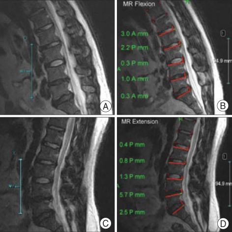
Methods: Segmental motion, defined as excessive (more than 3 mm) translational motion from flexion to extension, was investigated in 309 subjects (927 segments) using Kinetic MRI. Radiographic parameters which can help indicate segmental instability include disc degeneration (DD), facet joint osteoarthritis (FJO), and ligament flavum hypertrophy (LFH). These three radiographic parameters were simultaneously evaluated, and the combinations corresponding to significant segmental instability at each level were determined.
Results: The overall incidence of segmental instability was 10.5% at L3-L4, 16.5% at L4-L5, and 7.3% at L5-S1. DD and LFH at L3-L4 and FJO and LFH at L4-L5 were individually associated with segmental instability (p<0.05). At L4-L5, the following combinations had a higher incidence of segmental instability (p<0.05) when compared to other segments : (1) Grade IV DD with grade 3 FJO, (2) Grade 2 or 3 FJO with the presence of LFH, and (3) Grade IV DD with the presence of LFH. At L5-S1, the group with Grade III disc and Grade 3 FJO had a higher incidence of segmental instability than the group with Grade I or II DD and Grade 1 FJO.
Conclusion: This study showed that the presences of either Grade IV DD or grade 3 FJO with LFH at L4-L5 were good indicators for segmental instability. Therefore, using these parameters simultaneously in patients with segmental instability would be useful for determining candidacy for surgical treatment.
Keywords: Disc degeneration; Facet joint osteoarthritis; Hypertrophy of ligament flavum; MRI; Segmental instability.
Figures
References
-
- Berne D, Goubier JN, Lemoine J, Saillant G. The aging of the spine : natural evolution. Eur J Orthop Surg Traumatol. 1999;9:125–133.
-
- Boden SD, Davis DO, Dina TS, Patronas NJ, Wiesel SW. Abnormal magnetic-resonance scans of the lumbar spine in asymptomatic subjects. J Bone Joint Surg AM. 1990;72:403–408. - PubMed
-
- Edmondston SJ, Song S, Bricknell RV, Davies PA, Fersum K, Humphries P, et al. MRI evaluation of lumbar spine flexion and extension in asymptomatic individuals. Man Ther. 2000;5:158–164. - PubMed
-
- Elfering A, Semmer N, Birkhofer D, Zanetti M, Hodler J, Boos N. Risk factors for lumbar disc degeneration. Spine. 2002;27:125–134. - PubMed
LinkOut - more resources
Full Text Sources
