

Moyamoya-like vasculopathy in neurosarcoidosis
- PMID: 19242573
- PMCID: PMC2640829
- DOI: 10.3340/jkns.2009.45.1.50
Moyamoya-like vasculopathy in neurosarcoidosis
Abstract
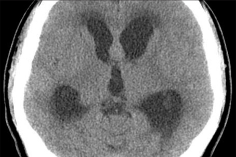
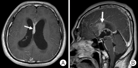
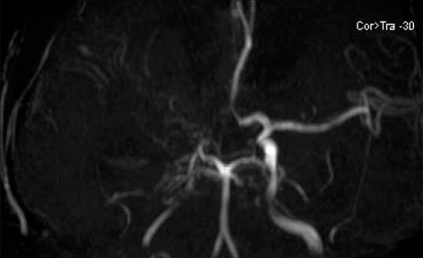
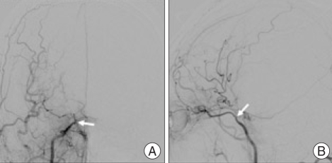
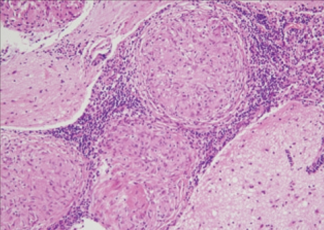
A 31-year-old man presented with dull headache and memory disturbance lasting for one week. Computed tomographic scans revealed acute hydrocephalus. The cerebrospinal fluid contained 53 leukocytes/mm(3), with a mononuclear preponderance and no erythrocytes. Magnetic resonance imaging revealed hydrocephalus and leptomeningeal enhancement. Magnetic resonance angiography and digital subtraction angiography showed supraclinoid occlusion of the right internal carotid artery, which resembled unilateral moyamoya disease. Neuroendoscopic biopsy of a lesion in the septum pellucidum revealed noncaseating granulomas, which was consistent with sarcoidosis. The patient was successfully managed with intravenous methylprednisolone and ventriculoperitoneal shunting. To our knowledge, this is the first case of moyamoya-like vasculopathy associated with neurosarcoidosis.
Keywords: Hydrocephalus; Moyamoya disease; Neurosarcoidosis.
Figures
References
-
- Benzagmout M, Boujraf S, Góngora-Rivera F, Bresson D, Van-Effenterre R. Neurosarcoidosis which manifested as acute hydrocephalus : diagnosis and treatment. Intern Med. 2007;46:1601–1604. - PubMed
-
- Brisman JL, Hinduja A, McKinney JS, Gerhardstein B. Successful emergent angioplasty of neurosarcoid vasculitis presenting with strokes. Surg Neurol. 2006;66:402–404. - PubMed
-
- Brown MM, Thompson AJ, Wedzicha JA, Swash M. Sarcoidosis presenting with stroke. Stroke. 1989;20:400–405. - PubMed
-
- Das SK, Sinha I, Kundu TN, Sanyal K, Santosh V, Shankar SK. Two cases of neurosarcoidosis presenting as peripheral neuropathy and stroke in young. J Assoc Physicians India. 1998;46:479–481. - PubMed
-
- Duffey P, Bates D. Transient focal neurological deficit in sarcoidosis. Sarcoidosis Vasc Diffuse Lung Dis. 1997;14:171–172. - PubMed
Publication types
LinkOut - more resources
Full Text Sources
