

In vivo investigation of breast cancer progression by use of an internal control
- PMID: 19242603
- PMCID: PMC2647724
- DOI: 10.1593/neo.08648
In vivo investigation of breast cancer progression by use of an internal control
Abstract
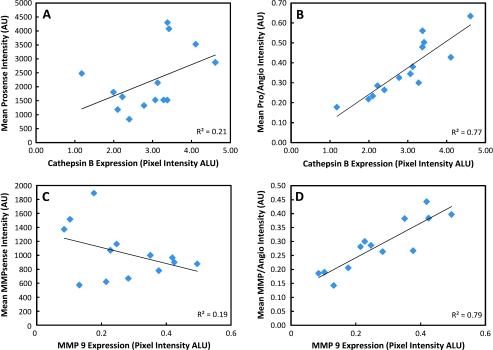
Optical imaging of breast cancer has been considered for detecting functional and molecular characteristics of diseases in clinical and preclinical settings. Applied to laboratory research, photonic investigations offer a highly versatile tool for preclinical imaging and drug discovery. A particular advantage of the optical method is the availability of multiple spectral bands for performing imaging. Herein, we capitalize on this feature to demonstrate how it is possible to use different wavelengths to offer internal controls and significantly improve the observation accuracy in molecular imaging applications. In particular, we show the independent in vivo detection of cysteine proteases along with tumor permeability and interstitial volume measurements using a dual-wavelength approach. To generate results with a view toward clinically geared studies, a transgenic Her2/neu mouse model that spontaneously developed mammary tumors was used. In vivo findings were validated against conventional ex vivo tests such as histology and Western blot analyses. By correcting for biodistribution parameters, the dual-wavelength method increases the accuracy of molecular observations by separating true molecular target from probe biodistribution. As such, the method is highly appropriate for molecular imaging studies where often probe delivery and target presence are not independently assessed. On the basis of these findings, we propose the dual-wavelength/normalization approach as an essential method for drug discovery and preclinical imaging studies.
Figures
References
-
- Barrett T, Koyama Y, Hama Y, Ravizzini G, Shin IS, Jang B-S, Paik CH, Choyke PL, Kobayashi H. Molecular imaging for typing epidermal growth factor receptors expressed on tumor cells using a cocktail of two monoclonal antibodies conjugated with two distinct near infrared fluorophores. Clin Cancer Res. 2007;15:6639–6648. - PubMed
-
- Montet X, Ntziachristos V, Grimm J, Weissleder R. Tomographic fluorescence mapping of tumor targets. Cancer Res. 2005;15:6330–6336. - PubMed
-
- Bremer C, Ntziachristos V, Weitkamp B, Theilmeier G, Heindel W, Weissleder R. Optical imaging of spontaneous breast tumors using protease sensing “smart” optical probes. Invest Radiol. 2005;40:321–327. - PubMed
-
- Deguchi JO, Aikawa M, Tung CH, Aikawa E, Kim DE, Ntziachristos V, Weissleder R, Libby P. Inflammation in atherosclerosis: visualizing matrix metalloproteinase action in macrophages in vivo. Circulation. 2006;114:55–62. - PubMed
Publication types
MeSH terms
Grants and funding
LinkOut - more resources
Full Text Sources
Medical
Research Materials
Miscellaneous