

Monocyte chemotactic protein 1 promotes lung cancer-induced bone resorptive lesions in vivo
- PMID: 19242604
- PMCID: PMC2647725
- DOI: 10.1593/neo.81282
Monocyte chemotactic protein 1 promotes lung cancer-induced bone resorptive lesions in vivo
Abstract
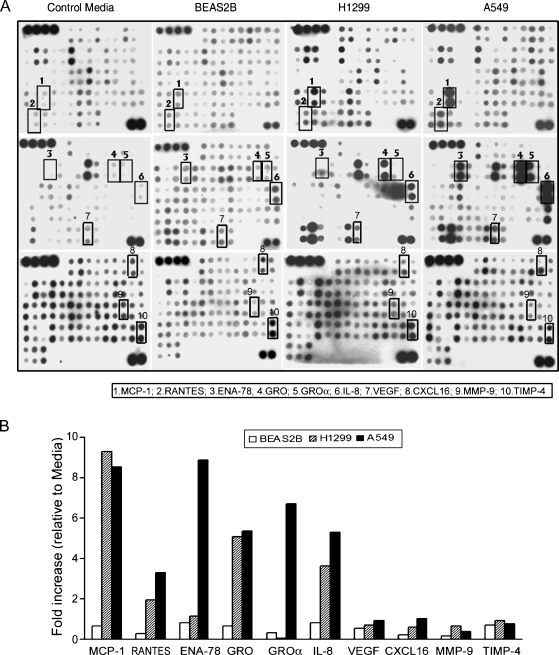
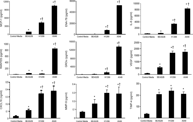
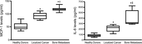
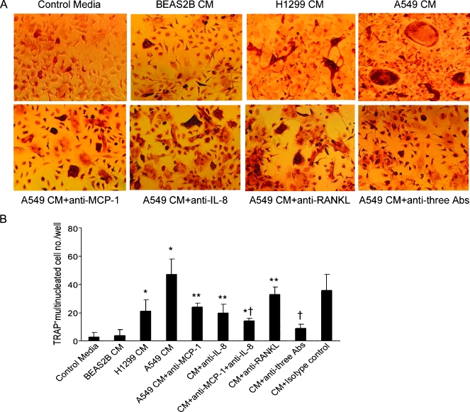
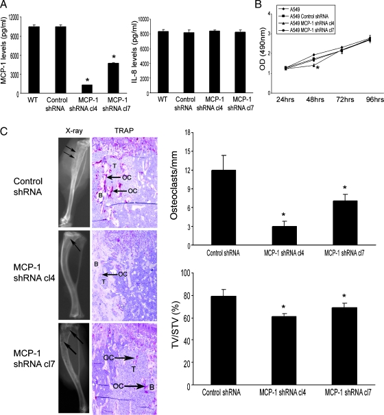
Lung cancer is the leading cause of cancer-related deaths. The morbidity and mortality of lung cancer have markedly increased in the past decade with at least 75% of patients with lung cancer having evidence of metastases at the time of diagnosis. It frequently metastasizes to bone resulting in osteolytic lesions with unknown mechanisms. The aim of this study was to identify factors that mediate lung cancer-induced osteoclast activity in vivo. Using a human cytokine antibody array, we first determined cytokine levels in a conditioned medium collected from non-small cell lung cancer A549 and H1299 cells and the non-neoplastic human bronchial epithelial BEAS2B cells. Both A549 and H1229 cells produced significantly higher amount of several cytokines including monocyte chemotactic protein 1 (MCP-1) and interleukin 8 (IL-8) compared with BEAS2B cells. These findings were confirmed by ELISA. From clinical serum specimens, we also observed that MCP-1 and IL-8 levels were increased in lung cancer patients with bone metastases compared with the patients with localized tumor. Next, we investigated the effects of MCP-1 on osteoclast formation in vitro using murine bone marrow-derived monocytes. A549 conditioned medium induced osteoclast formation that was inhibited by neutralizing antibodies against MCP-1. Finally, A549 cells were stably transfected with MCP-1 short hairpin RNA. The MCP-1 knockdown A549 cells were implanted into the tibia of severe combined immunodeficient mice for 4 weeks. The MCP-1 knockdown significantly diminished A549 cell growth. We conclude that MCP-1 promotes lung cancer-induced osteoclast activity and thus bone resorptive lesions in vivo.
Figures
Similar articles
-
Activation of MCP-1/CCR2 axis promotes prostate cancer growth in bone.Clin Exp Metastasis. 2009;26(2):161-9. doi: 10.1007/s10585-008-9226-7. Epub 2008 Nov 11. Clin Exp Metastasis. 2009. PMID: 19002595
-
Monocyte chemotactic protein-1 mediates prostate cancer-induced bone resorption.Cancer Res. 2007 Apr 15;67(8):3646-53. doi: 10.1158/0008-5472.CAN-06-1210. Cancer Res. 2007. PMID: 17440076
-
Mixed metastatic lung cancer lesions in bone are inhibited by noggin overexpression and Rank:Fc administration.J Bone Miner Res. 2006 Oct;21(10):1571-80. doi: 10.1359/jbmr.060706. J Bone Miner Res. 2006. PMID: 16995812
-
Tumor-derived interleukin-8 stimulates osteolysis independent of the receptor activator of nuclear factor-kappaB ligand pathway.Cancer Res. 2005 Dec 1;65(23):11001-9. doi: 10.1158/0008-5472.CAN-05-2630. Cancer Res. 2005. PMID: 16322249
-
Monocyte Chemoattractant Protein-1 (MCP-1/CCL2) Drives Activation of Bone Remodelling and Skeletal Metastasis.Curr Osteoporos Rep. 2019 Dec;17(6):538-547. doi: 10.1007/s11914-019-00545-7. Curr Osteoporos Rep. 2019. PMID: 31713180 Free PMC article. Review.
Cited by
-
Surgical removal of the parametrial fat pads stimulates apoptosis and inhibits UVB-induced carcinogenesis in mice fed a high-fat diet.Proc Natl Acad Sci U S A. 2012 Jun 5;109(23):9065-70. doi: 10.1073/pnas.1205810109. Epub 2012 May 21. Proc Natl Acad Sci U S A. 2012. PMID: 22615388 Free PMC article.
-
The effect of down regulation of calcineurin Aα by lentiviral vector-mediated RNAi on the biological behavior of small-cell lung cancer and its bone metastasis.Clin Exp Metastasis. 2011 Dec;28(8):765-78. doi: 10.1007/s10585-011-9408-6. Epub 2011 Jul 23. Clin Exp Metastasis. 2011. PMID: 21785830
-
Immune crosstalk in cancer progression and metastatic spread: a complex conversation.Nat Rev Immunol. 2020 Aug;20(8):483-497. doi: 10.1038/s41577-019-0271-z. Epub 2020 Feb 5. Nat Rev Immunol. 2020. PMID: 32024984 Review.
-
CC Chemokines in a Tumor: A Review of Pro-Cancer and Anti-Cancer Properties of the Ligands of Receptors CCR1, CCR2, CCR3, and CCR4.Int J Mol Sci. 2020 Nov 9;21(21):8412. doi: 10.3390/ijms21218412. Int J Mol Sci. 2020. PMID: 33182504 Free PMC article. Review.
-
Modulating the tumor microenvironment: The role of traditional Chinese medicine in improving lung cancer treatment.Open Life Sci. 2025 May 20;20(1):20251100. doi: 10.1515/biol-2025-1100. eCollection 2025. Open Life Sci. 2025. PMID: 40417000 Free PMC article. Review.
References
-
- Jemal A, Siegel R, Ward E, Hao Y, Xu J, Murray T, Thun MJ. Cancer statistics, 2008. CA Cancer J Clin. 2008;58:71–96. - PubMed
-
- Mulshine JL, Sullivan DC. Clinical practice. Lung cancer screening. N Engl J Med. 2005;352:2714–2720. - PubMed
-
- Keller ET, Dai J, Escara-Wilke J, Hall CL, Ignatoski K, Taichman RS, Keller J. New trends in the treatment of bone metastasis. J Cell Biochem. 2007;102:1095–1102. - PubMed
-
- Becker S, Quay J, Koren HS, Haskill JS. Constitutive and stimulated MCP-1, GRO alpha, beta, and gamma expression in human airway epithelium and bronchoalveolar macrophages. Am J Physiol. 1994;266:L278–L286. - PubMed
Publication types
MeSH terms
Substances
Grants and funding
LinkOut - more resources
Full Text Sources
Other Literature Sources
Medical
Research Materials
Miscellaneous