. 2009 Sep;467(9):2403-13.
doi: 10.1007/s11999-009-0762-5. Epub 2009 Feb 26.

Differences between sagittal femoral mechanical and distal reference axes should be considered in navigated TKA

Affiliations

Differences between sagittal femoral mechanical and distal reference axes should be considered in navigated TKA

Byung June Chung et al. Clin Orthop Relat Res. 2009 Sep.

Abstract

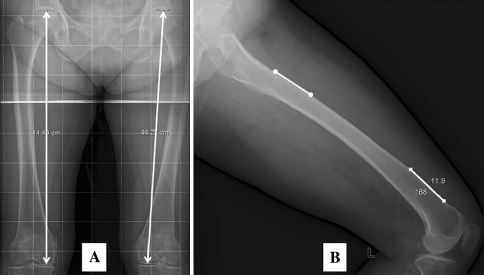
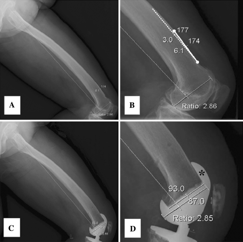
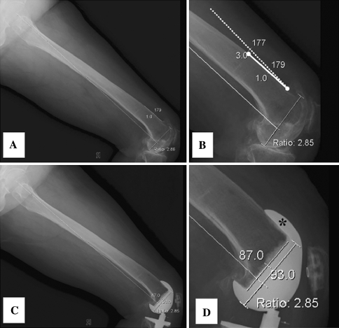
In computer-assisted TKA, surgeons determine positioning of the femoral component in the sagittal plane based on the sagittal mechanical axis identified by the navigation system. We hypothesized mechanical and distal femoral axes may differ on lateral views and these variations are influenced by anteroposterior bowing and length of the femur. We measured angles between the mechanical axis and distal femoral axis on 200 true lateral radiographs of the whole femur from 100 adults. We used multivariate linear regression to identify predictors of differences between the axes. Depending on the method used to define the two axes, the mean angular difference between the axes was as much as 3.8 degrees and as little as 0.0 degrees, with standard differences ranging from 1.7 degrees to 1.9 degrees. Variation between the two axes increased with increased femoral bowing and increased femoral length. Surgeons should consider differences between the mechanical axes and distal femoral axes when they set the sagittal plane position of a femoral component in navigated cases. Our findings also may be relevant when measuring rotation of the femoral component in the sagittal plane from postoperative radiographs or when interpreting femoral component sagittal rotation results reported in other studies.


Figures

Fig. 1
Fig. 1
To obtain a true lateral view of the whole femur, patients placed their thigh on a 17-inch × 17-inch digital flat detector in a diagonal position. The xray beam tube was tilted 15° to directly aim the center of the guide beam toward the center of the digital detector (solid arrow) and the midpoint of the patient’s thigh (asterisk). A sponge block (open arrow) was placed to hold the patient’s leg in a right position.
Fig. 2A–C
Fig. 2A–C
(A) A radiograph shows how mechanical axis 1 was defined. (B) An intraoperative photograph and (C) a radiograph display the registration point (asterisk) for the distal femur center and the top point of the intercondylar notch (solid arrow).
Fig. 3A–B
Fig. 3A–B
(A) A schematic drawing shows how the distal femur center is identified by the OrthoPilot® system. (B) A lateral radiograph shows mechanical axis 2 was defined to represent the point 65% posteriorly in the connecting line between the anterior cortical line and the most prominent point of the posterior femoral condyle.
Fig. 4A–D
Fig. 4A–D
The radiographic measurements of the differences are shown between (A) mechanical axis 1 versus the distal anterior cortex axis, (B) mechanical axis 1 versus the distal medullary axis, (C) mechanical axis 2 versus the distal anterior cortex axis, and (D) mechanical axis 2 versus the distal medullary axis.
Fig. 5A–B
Fig. 5A–B
(A) The femur length was measured on long cassette AP radiographs using grids, and (B) the degrees of bowing were determined by measuring the angle between the anterior cortical line and the proximal cortical line.
Fig. 6A–D
Fig. 6A–D
(A) A preoperative radiographic assessment of a patient shows the anterior cortical line is oriented in a position of 6° flexion relative to the mechanical axis 2. (B) Projected into the anterior cortex are the line (dotted line) oriented in the position of 3° flexion reflecting the design feature of 3° flexion in the anterior flange of a femoral component. (C) A postoperative radiograph shows distal femur resection was performed at an angle of 3° flexion relative to the mechanical axis 2. (D) The anterior flange of the femoral component (asterisk) sits perfectly on the anterior cortical surface without anterior notching or anterior flange prominence.
Fig. 7A–D
Fig. 7A–D
(A) A preoperative radiographic assessment of a patient shows the anterior cortical line is oriented in a position of 1° extension with respect to the mechanical axis 2. (B) Projected off the anterior cortex is the line (dotted line) oriented in the position of 3° reflecting the design feature of 3° flexion in the anterior flange of a femoral component. (C) A postoperative radiograph shows distal femur resection was performed at an angle of 3° extension relative to the mechanical axis 2. (D) The anterior flange of the femoral component (asterisk) sits perfectly on the anterior cortical surface without anterior notching or anterior flange prominence.

References

    1. Anderson KC, Buehler KC, Markel DC. Computer assisted navigation in total knee arthroplasty: comparison with conventional methods. J Arthroplasty. 2005;20:132–138. doi: 10.1016/j.arth.2005.05.009. - DOI - PubMed
    1. Bathis H, Perlick L, Tingart M, Luring C, Zurakowski D, Grifka J. Alignment in total knee arthroplasty: a comparison of computer-assisted surgery with the conventional technique. J Bone Joint Surg Br. 2004;86:682–687. doi: 10.1302/0301-620X.86B5.14927. - DOI - PubMed
    1. Bolognesi M, Hofmann A. Computer navigation versus standard instrumentation for TKA: a single-surgeon experience. Clin Orthop Relat Res. 2005;440:162–169. doi: 10.1097/01.blo.0000186561.70566.95. - DOI - PubMed
    1. Brattstrom H. Patella alta in non-dislocating knee joints. Acta Orthop Scand. 1970;41:578–588. - PubMed
    1. Chauhan SK, Scott RG, Breidahl W. Beaver RJ: Computer-assisted knee arthroplasty versus a conventional jig-based technique: a randomised, prospective trial. J Bone Joint Surg Br. 2004;86:372–377. doi: 10.1302/0301-620X.86B3.14643. - DOI - PubMed

Publication types