

Mechanical stimulation alters tissue differentiation and molecular expression during bone healing
- PMID: 19242967
- PMCID: PMC2726267
- DOI: 10.1002/jor.20863
Mechanical stimulation alters tissue differentiation and molecular expression during bone healing
Abstract
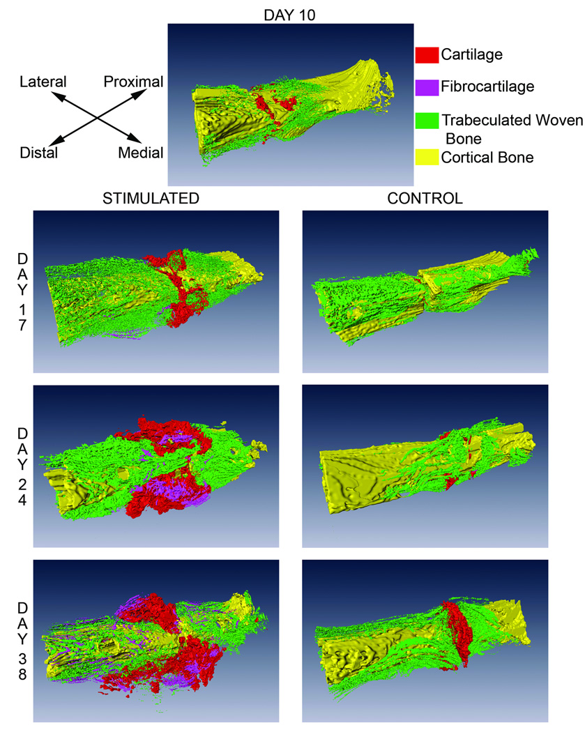
Further understanding of how mechanical cues modulate skeletal tissue differentiation can identify potential means of enhancing repair following injury or disease. Prior studies examined the effects of mechanical loading on osteogenesis, chondrogenesis, and fibrogenesis in an effort to enhance bony union. However, exploring how mechanical stimuli can divert the bone healing process towards formation of other mesenchymal tissues, as an endpoint, may elucidate new avenues for repair and regeneration of tissues such as cartilage and fibrous tissue. This study investigated the use of mechanical stimulation to promote cartilage rather than bone formation within an osteotomy. Our overall goal was to define skeletal tissue distribution and molecular expression patterns induced by the stimulation. Retired breeder Sprague-Dawley rats (n = 85) underwent production of a mid-diaphyseal, transverse femoral osteotomy followed by external fixation. Beginning on postoperative day 10 and continuing for 1, 2, or 4 weeks, a cyclic bending motion (+35 degrees/-25 degrees at 1 Hz) was applied in the sagittal plane for 15 min/day for 5 consecutive days/week. Control animals experienced continuous rigid fixation. Histological and molecular analyses indicated that stimulation substantially altered normal bone healing. Stimulated specimens exhibited an increase in cartilage volume over time, while control specimens demonstrated bony bridging. Stimulation induced upregulation of cartilage-related genes (COL2A1 and COL10A1) and downregulation of bone morphogenetic proteins (BMPs) -4, -6 and -7. However, BMP-3 was upregulated with stimulation. These findings illustrate that mechanical cues can selectively modulate osteogenesis and chondrogenesis in vivo, and suggest a potential basis for treatment regimens for injured or diseased cartilaginous tissues.
(c) 2009 Orthopaedic Research Society.
Figures


References
-
- Dowthwaite GP, Flannery CR, Flannelly J, et al. A mechanism underlying the movement requirement for synovial joint cavitation. Matrix Biol. 2003;22(4):311–322. - PubMed
-
- Vortkamp A, Pathi S, Peretti GM, et al. Recapitulation of signals regulating embryonic bone formation during postnatal growth and in fracture repair. Mech Dev. 1998;71(1–2):65–76. - PubMed
-
- Carter DR, Beaupre GS, Giori NJ, Helms JA. Mechanobiology of skeletal regeneration. Clin Orthop Relat Res. 1998;(355 Suppl):S41–S55. - PubMed
-
- Goodship AE, Cunningham JL, Kenwright J. Strain rate and timing of stimulation in mechanical modulation of fracture healing. Clin Orthop. 1998;(355 Suppl):S105–S115. - PubMed
-
- Le AX, Miclau T, Hu D, Helms JA. Molecular aspects of healing in stabilized and non-stabilized fractures. J Orthop Res. 2001;19(1):78–84. - PubMed
Publication types
MeSH terms
Substances
Grants and funding
LinkOut - more resources
Full Text Sources
Medical
Molecular Biology Databases
