

Review
doi: 10.1021/cr800470j.
Polyubiquitin binding and disassembly by deubiquitinating enzymes
Affiliations
- PMID: 19243136
- PMCID: PMC2734106
- DOI: 10.1021/cr800470j
Item in Clipboard
Review
Polyubiquitin binding and disassembly by deubiquitinating enzymes
Chem Rev.
2009 Apr.
No abstract available
Figures
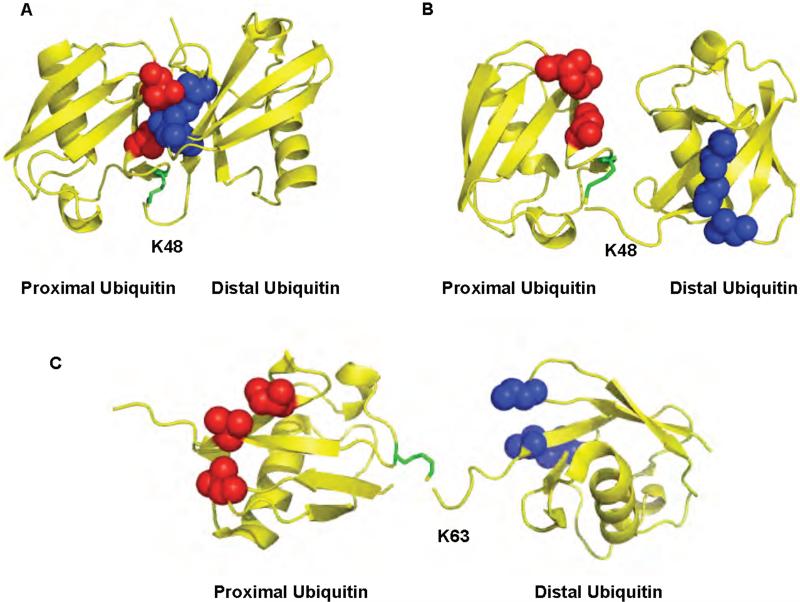
The structures of K48 and K63-linked diubiquitin. A. Closed conformation of K48-linked diubiquitin. B. Open conformation of K48-linked diubiquitin. C. Extended conformation of K63-linked diubiquitin. The ubiquitin moieties in the chain are colored in yellow. The hydrophobic patch in the distal and proximal ubiquitins is shown in blue and red respectively. K48 or K63 of ubiquitin that participated in the isopeptide bond is colored in green. The protein data bank codes are 1AAR, 1TBE, and 2JF5 for A, B, and C, respectively
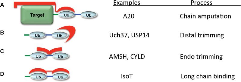
Possible mechanisms of polyubiquitin recognition by DUBs. A. DUBs can interact with both the target protein and polyubiquitin. B. DUBs can recognize of the distal ubiquitin the chain. C. DUBs could bind simultaneously two ubiquitins by interacting with surfaces on both ubiquitins that surround the isopeptide bond. D. Finally, a DUB can recognize polyubiquitin through the use of multiple ubiquitin binding domains. Polyubiquitin is shown in blue, the target protein in green, and the polyubiquitin receptor in red.
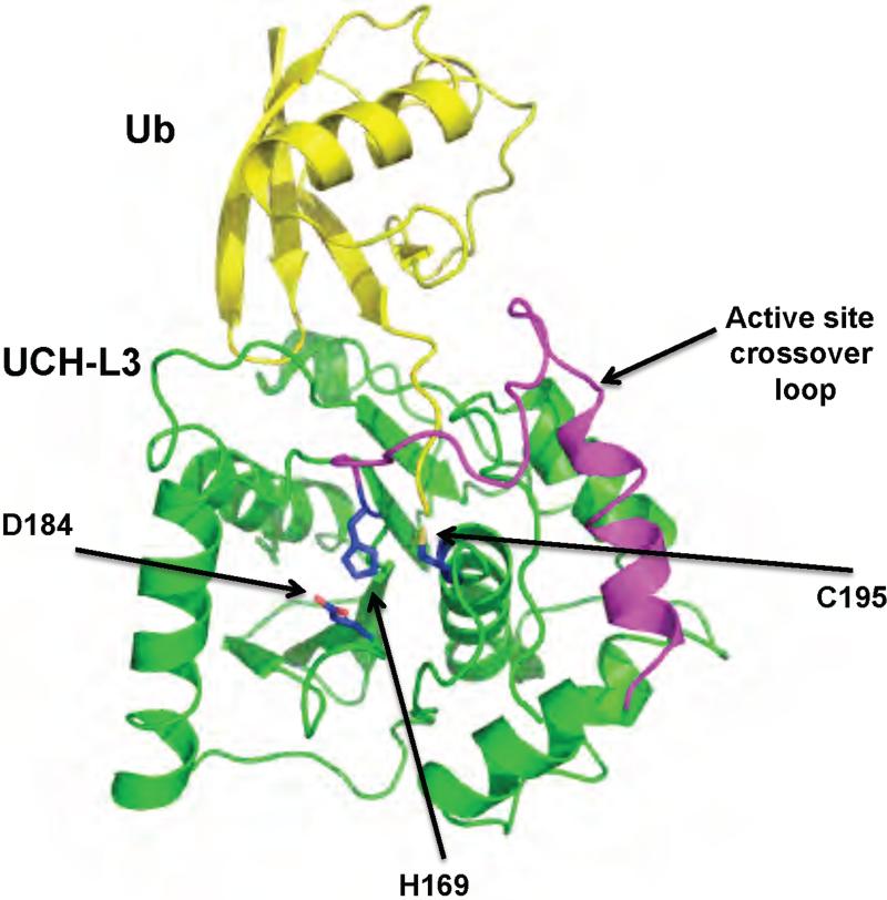
Structure of a UCH family DUB bound to Ub-VME, UCH-L3. Ub-VME is colored in yellow, and UCH-L3 in green. The active site loop is colored in magenta. The active site residues are shown in blue. The protein data bank identification code is 1XD3.
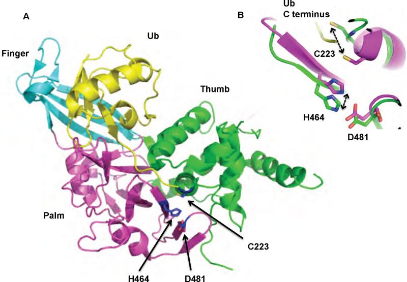
Structure of a UBP/USP family DUB, USP7. A. Structure of Usp7 bound to Ubal. Ubal is colored in yellow. The three domains that make up the USP domain are colored in cyan (Finger), green (Thumb), and magenta (Palm). The active site residues are shown in blue. B. Conformational rearrangement of the active site residues upon binding of Ubal to Usp7. Ubal unbound and bound Usp7 are colored in green and magenta respectively. The ubiquitin C terminus is colored in yellow. The protein data bank identification codes for the unbound and bound structure of Usp7 are 1NB8 and 1NBF respectively.
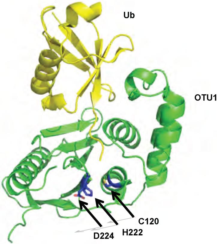
Structure of yeast OTU1 bound to Ub-Br3. OTU1 is shown in green. Ub-Br3 is shown in yellow. The active site residues are shown in blue. The protein data bank identification code is 3BY4.
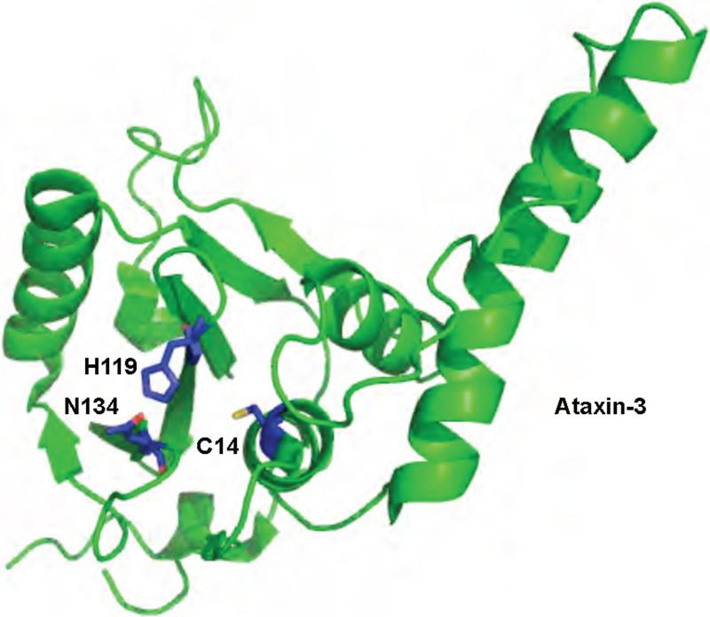
Structure of a Josephin domain family DUB, Ataxin-3. Ataxin-3 is shown in green. The active site residues are shown in blue. The protein data bank identification code is 1YZB.
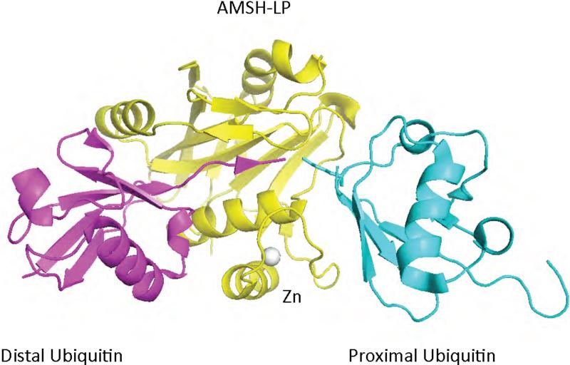
Structure of a JAMM domain (yellow) from the human AMSH-like protein complexed to K63-linked di-ubiquitin (cyan and magenta). The structure consists of the JAMM domain core surrounding the zinc atom (grey) and two AMSH-specific inserts that interact with the proximal (cyan) and distal (magenta) domains of diubiquitin. The protein data bank identification code is 2ZNV.
References
Publication types
MeSH terms
Substances
Grants and funding
LinkOut - more resources
Full Text Sources
