

Imaging pancreatic cancer using bioconjugated InP quantum dots
- PMID: 19243145
- PMCID: PMC2762404
- DOI: 10.1021/nn8008933
Imaging pancreatic cancer using bioconjugated InP quantum dots
Abstract
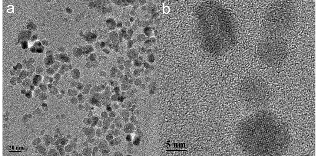
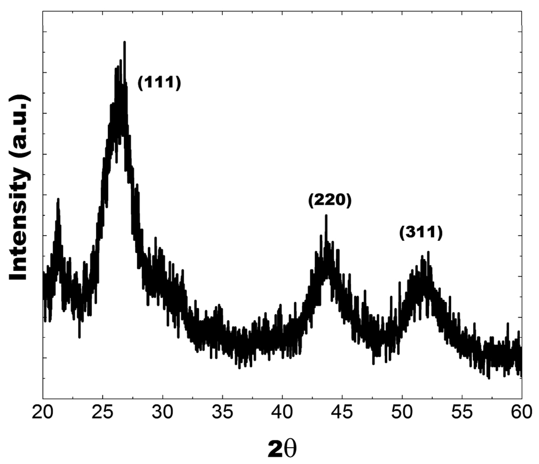
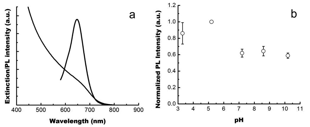
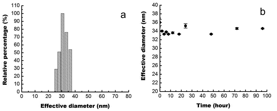
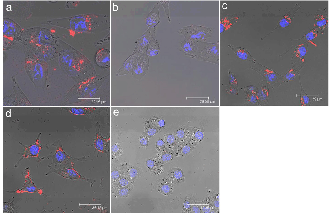
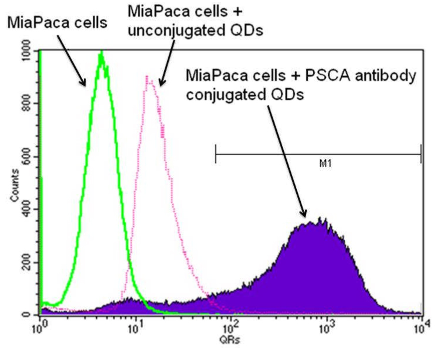
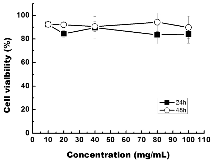
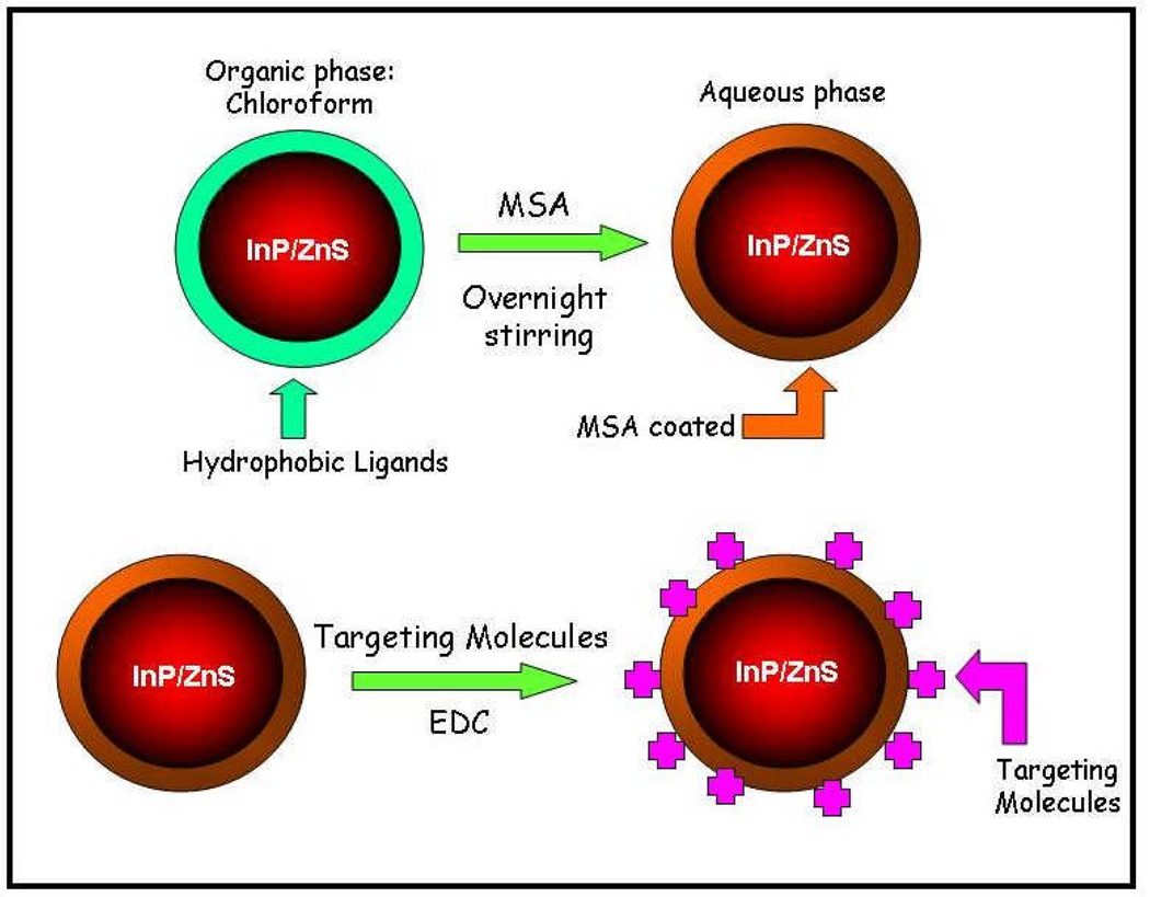
In this paper, we report the successful use of non-cadmium-based quantum dots (QDs) as highly efficient and nontoxic optical probes for imaging live pancreatic cancer cells. Indium phosphide (core)-zinc sulfide (shell), or InP/ZnS, QDs with high quality and bright luminescence were prepared by a hot colloidal synthesis method in nonaqueous media. The surfaces of these QDs were then functionalized with mercaptosuccinic acid to make them highly dispersible in aqueous media. Further bioconjugation with pancreatic cancer specific monoclonal antibodies, such as anticlaudin 4 and antiprostate stem cell antigen (anti-PSCA), to the functionalized InP/ZnS QDs, allowed specific in vitro targeting of pancreatic cancer cell lines (both immortalized and low passage ones). The receptor-mediated delivery of the bioconjugates was further confirmed by the observation of poor in vitro targeting in nonpancreatic cancer based cell lines which are negative for the claudin-4-receptor. These observations suggest the immense potential of InP/ZnS QDs as non-cadmium-based safe and efficient optical imaging nanoprobes in diagnostic imaging, particularly for early detection of cancer.
Figures
References
-
- Prasad PN. Nanophotonics. Hoboken, NJ: Wiley; 2004.
-
- Prasad PN. Introduction to Biophotonics. Hoboken, NJ: Wiley-Interscience; 2003.
-
- Arya H, Kaul Z, Wadhwa R, Taira K, Hirano T, Kaul SC. Quantum Dots in Bio-imaging: Revolution by the Small. Biochemical and Biophysical Research Communications. 2005;329:1173–1177. - PubMed
-
- Azzazy HME, Mansour MMH, Kazmierczak SC. From Diagnostics to Therapy: Prospects of Quantum Dots. Clinical Biochemistry. 2007;40:917–927. - PubMed
-
- Bruchez M, Jr, Moronne M, Gin P, Weiss S, Alivisatos AP. Semiconductor Nanocrystals as Fluorescent Biological Labels. Science. 1998;281:2013–2016. - PubMed
Publication types
MeSH terms
Substances
Grants and funding
LinkOut - more resources
Full Text Sources
Other Literature Sources
Medical
