

Relevance of biophysical interactions of nanoparticles with a model membrane in predicting cellular uptake: study with TAT peptide-conjugated nanoparticles
- PMID: 19243206
- PMCID: PMC2757462
- DOI: 10.1021/mp900011h
Relevance of biophysical interactions of nanoparticles with a model membrane in predicting cellular uptake: study with TAT peptide-conjugated nanoparticles
Abstract
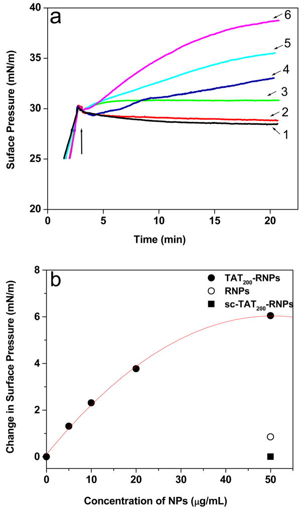
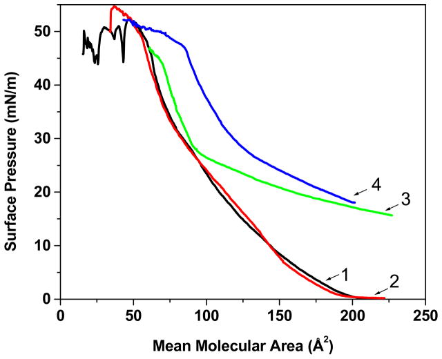
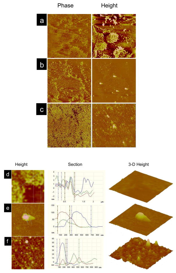
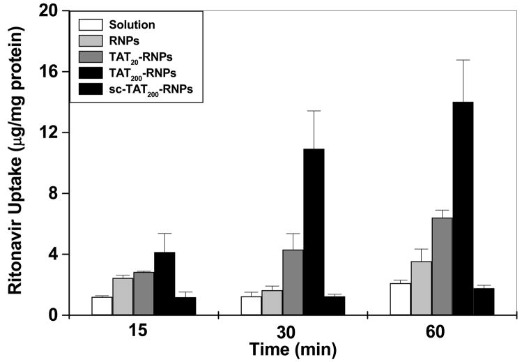
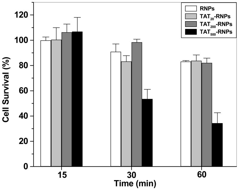
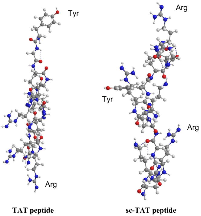
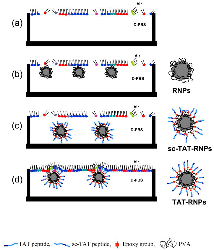
The aim of the study was to test the hypothesis that the biophysical interactions of the trans-activating transcriptor (TAT) peptide-conjugated nanoparticles (NPs) with a model cell membrane could predict the cellular uptake of the encapsulated therapeutic agent. To test the above hypothesis, the biophysical interactions of ritonavir-loaded poly(l-lactide) nanoparticles (RNPs), conjugated to either a TAT peptide (TAT-RNPs) or a scrambled TAT peptide (sc-TAT-RNPs), were studied with an endothelial cell model membrane (EMM) using a Langmuir film balance, and the corresponding human vascular endothelial cells (HUVECs) were used to study the uptake of the encapsulated therapeutic. Biophysical interactions were determined from the changes in surface pressure (SP) of the EMM as a function of time following interaction with NPs, and the compression isotherm (pi-A) of the EMM lipid mixture in the presence of NPs. In addition, the EMMs were transferred onto a silicon substrate following interactions with NPs using the Langmuir-Schaeffer (LS) technique. The transferred LS films were imaged by atomic force microscopy (AFM) to determine the changes in lipid morphology and to characterize the NP-membrane interactions. TAT-RNPs showed an increase in SP of the EMM, which was dependent upon the amount of the peptide bound to NPs and the concentration of NPs, whereas sc-TAT-RNPs and RNPs did not show any significant change in SP. The isotherm experiment showed a shift toward higher mean molecular area (mmA) in the presence of TAT-RNPs, indicating their interactions with the lipids of the EMM, whereas sc-TAT-RNPs and RNPs did not show any significant change. The AFM images showed condensation of the lipids following interaction with TAT-RNPs, indicating their penetration into the EMM, whereas RNPs did not cause any change. Surface analysis and 3-D AFM images of the EMM further confirmed penetration of TAT-RNPs into the EMM, whereas RNPs were seen anchored loosely to the membrane, and were significantly less in number than TAT-RNPs. We speculate that hydrophobic tyrosine of the TAT that forms the NP-interface drives the initial interactions of TAT-RNPs with the EMM, followed by electrostatic interactions with the anionic phospholipids of the membrane. In the case of sc-TAT-RNPs, hydrophilic arginine forms the NP-interface that does not interact with the EMM, despite having the similar cationic charge on these NPs as TAT-RNPs. TAT peptide alone did not show any change in SP, suggesting that the interaction occurs when the peptide is conjugated to a carrier system. HUVECs showed higher uptake of the drug with TAT-RNPs as compared to that with sc-TAT-RNPs or RNPs, suggesting that the biophysical interactions of NPs with cell membrane lipids play a role in cellular internalization of NPs. In conclusion, TAT peptide sequence and the amount of TAT conjugated to NPs significantly affect the biophysical interactions of NPs with the EMM, and these interactions correlate with the cellular delivery of the encapsulated drug. Biophysical interactions with a model membrane thus could be effectively used in developing efficient functionalized nanocarrier systems for drug delivery applications.
Figures
References
-
- Deshayes S, Simeoni F, Morris MC, Divita G, Heitz F. Peptide-mediated delivery of nucleic acids into mammalian cells. Methods Mol Biol. 2007;386:299–308. - PubMed
-
- Deshayes S, Morris M, Heitz F, Divita G. Delivery of proteins and nucleic acids using a non-covalent peptide-based strategy. Adv Drug Del Rev. 2008;60:537–547. - PubMed
-
- Panyam J, Labhasetwar V. Dynamics of endocytosis and exocytosis of poly(D,L-lactide-co-glycolide) nanoparticles in vascular smooth muscle cells. Pharm Res. 2003;20:212–220. - PubMed
-
- Trehin R, Merkle HP. Chances and pitfalls of cell penetrating peptides for cellular drug delivery. Eur J Pharm Biopharm. 2004;58:209–223. - PubMed
Publication types
MeSH terms
Substances
Grants and funding
LinkOut - more resources
Full Text Sources
Research Materials
Miscellaneous
