. 2010 Jan;20(1):211-21.
doi: 10.1111/j.1750-3639.2009.00269.x. Epub 2009 Feb 20.

A2B5 cells from human glioblastoma have cancer stem cell properties

Affiliations

A2B5 cells from human glioblastoma have cancer stem cell properties

Aurélie Tchoghandjian et al. Brain Pathol. 2010 Jan.

Abstract

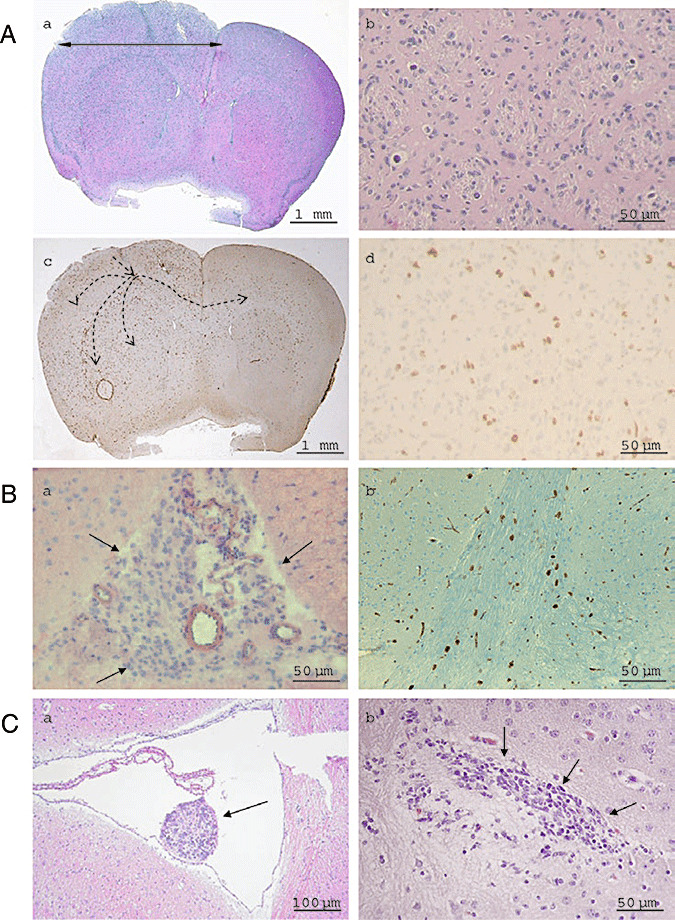
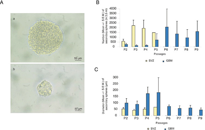
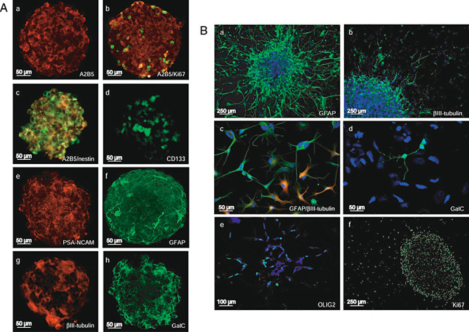
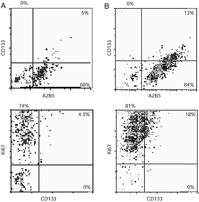
Glioblastomas, like other cancers, harbor small cell populations with the capability of sustaining tumor formation. These cells are referred to as cancer stem cells. We isolated cells expressing the surface marker A2B5 from three human glioblastomas (GBM) and showed that after grafting into nude mice, they generated dense and highly infiltrative tumors. Then, we extensively studied A2B5(+) cells isolated from 11 human GBM. These cells display neurosphere-like, self-renewal, asymmetrical cell division properties and have multipotency capability. Stereotactic xenografts of dissociated A2B5(+)-derived secondary spheres revealed that as few as 1000 cells produced a tumor. Moreover, flow cytometry characterization of A2B5(+)-derived spheres revealed three distinct populations of cells: A2B5(+)/CD133(+), A2B5(+)/CD133(-) and A2B5(-)/CD133(-), with striking proportion differences among GBM. Both A2B5(+)/CD133(+) and A2B5(+)/CD133(-) cell fractions displayed a high proliferative index, the potential to generate spheres and produced tumors in nude mice. Finally, we generated two green fluorescent protein-cell lines that display--after serum induction--distinct proliferative and migratory properties, and differ in their CD133 level of expression. Taken together, our results suggest that transformed A2B5(+) cells are crucial for the initiation and maintenance of GBM, although CD133 expression is more involved in determining the tumor's behavior.


Figures

Figure 1
Figure 1
A. A2B5+ cells are tumorigenic after transplantation in nude mice brain: hematoxylin‐eosin histology (a, b) and anti‐Ki67 immunohistochemistry (c, d). Dotted arrows illustrate injected cells crossing the corpus callosum to colonize the contra‐lateral hemisphere (c). Dense tumors, made by polymorphic and highly mitotic cells (b) which infiltrate the whole hemisphere are observed, giving rise to a larger hemisphere (a). B. Secondary spheres obtained from cultured A2B5+ GBM cells are tumorigenic in vivo (GBM 6, 100 000 cells injected). Subarachnoidal spaces are filled with tumor (a). Tumor cells infiltrate the corpus callosum and express the proliferative marker Ki67 (b). C. Hematoxylin‐eosin histology of tumors obtained from injection of 1 000 cells A2B5+/CD133 (a) and 1000 cells A2B5+/CD133+ (b) isolated by flow cytometry from secondary spheres derived from GBM 6.
Figure 2
Figure 2
Persistence of self‐renewal and high proliferation capacity during passages for glioblastomas (GBM) secondary spheres derived from A2B5 + isolated cells compared with subventricular zone (SVZ). A. Phase contrast images of a sphere derived from GBM (a) and SVZ (b). B. Mean number of secondary spheres in 3.5 mL of stem‐cell permissive medium for 4 GBM (GBM 4, 5, 6 and 7) vs. 4 SVZ, showing a persistent self‐renewal capacity in GBM and a limited one in SVZ. C. Mean diameter of secondary spheres recorded in 4 GBM (GBM 4, 5, 6 and 7) compared with 4 SVZ. Mann–Whitney test showed that the diameter of GBM spheres is statistically higher than the diameter of SVZ spheres (P < 0.02). For B and C, results are expressed as mean ± standard error of the mean (SEM). Although bar error remains small for SVZ spheres, they are high for GBM spheres reflecting the heterogeneity of these tumors.
Figure 3
Figure 3
A. Neural stem cells (NSC) and differentiation markers in glioblastomas (GBM) secondary spheres cultured in stem cell‐permissive medium. (a) A2B5, (b) A2B5 (red)/Ki67 (green), (c) A2B5 (red)/nestin (green), (d) CD133, (e) polysialylated neural cell adhesion molecule (PSA‐NCAM), (f) glial fibrillary acid protein (GFAP), (g) βIII‐tubulin, (h) GalC. B. NSC and differentiation markers in GBM secondary spheres cultured in differentiation medium. (a) GFAP, (b) βIII‐tubulin, (c) GFAP (red)/βIII‐tubulin (green), (d) GalC, (e) OLIG2, (f) Ki67.
Figure 4
Figure 4
Flow cytometry analysis of secondary spheres derived from glioblastomas (GBM) A2B5+ cells A2B5, CD133 and Ki67 expression. A. GBM 6, passage 15: 5% of A2B5+/CD133+; 68% of A2B5+/CD133; 27% of A2B5/CD133. Similar results were obtained at passage 34. B. GBM 7, passage 13: 13% of A2B5+/CD133+; 84% of A2B5+/CD133; 3% of A2B5/CD133. All CD133+ cells express the Ki67 antigen but the major part of Ki67 expressing cells does not express CD133. Some CD133 cells do not proliferate (A,B).
Figure 5
Figure 5
Correlation between in vitro behavior of glioblastomas (GBM) 6 and GBM 9 EGFP cell lines, and Magnetic Resonance Imaging (MRI) images of the primary tumor of the corresponding patient. A. MRI scans show that GBM 6 tumor contacts lateral ventricle (left) and GBM 9 tumor is located in the cortex (right). B. GBM 6 and GBM 9 cell lines migration and proliferation rates. Pictures were obtained after acquisition at day 0 and at day 3 of monolayer culture using the flash‐cytometer. C. GBM 6 cells show the higher migration rate (left) and GBM 9 cells the higher proliferation rate (right). *P < 0.005. Abbreviation: SEM = standard error of the mean.

Similar articles

Cited by

References

    1. Bao S, Wu Q, McLendon RE, Hao Y, Shi Q, Hjelmeland AB et al (2006) Glioma stem cells promote radioresistance by preferential activation of the DNA damage response. Nature 444:756–760. - PubMed
    1. Beier D, Hau P, Proescholdt M, Lohmeier A, Wischhusen J, Oefner PJ et al (2007) CD133(+) and CD133(−) glioblastoma‐derived cancer stem cells show differential growth characteristics and molecular profiles. Cancer Res 67:4010–4015. - PubMed
    1. Calabrese C, Poppleton H, Kocak M, Hogg TL, Fuller C, Hamner B et al (2007) A perivascular niche for brain tumor stem cells. Cancer Cell 11:69–82. - PubMed
    1. Colin C, Baeza N, Tong S, Bouvier C, Quilichini B, Durbec P, Figarella‐Branger D (2006) In vitro identification and functional characterization of glial precursor cells in human gliomas. Neuropathol Appl Neurobiol 32:189–202. - PubMed
    1. Galli R, Binda E, Orfanelli U, Cipelletti B, Gritti A, De Vitis S et al (2004) Isolation and characterization of tumorigenic, stem‐like neural precursors from human glioblastomas. Cancer Res 64:7011–7021. - PubMed

Publication types

MeSH terms