. 2009 May;100(5):940-7.
doi: 10.1111/j.1349-7006.2009.01119.x. Epub 2009 Feb 19.

Knockdown of c-FLIP(L) enhanced AD5-10 anti-death receptor 5 monoclonal antibody-induced apoptosis in human lung cancer cells

Affiliations

Knockdown of c-FLIP(L) enhanced AD5-10 anti-death receptor 5 monoclonal antibody-induced apoptosis in human lung cancer cells

Feng Chen et al. Cancer Sci. 2009 May.

Erratum in

  • Cancer Sci. 2009 Dec;100(12):2469

Abstract

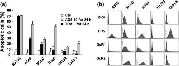
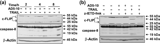
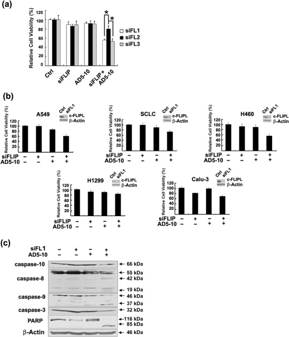
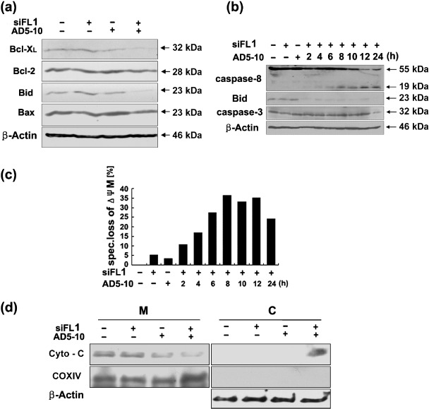
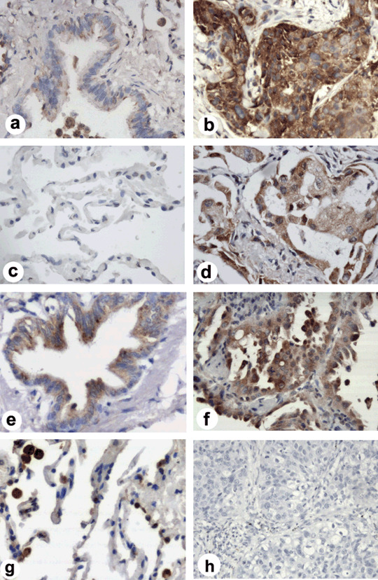
It is reported that the agonistic antibodies against death receptors 4 and 5 (DR4, DR5) are cytotoxic to various cancer cells. In the present study, the sensitivity of five human lung cancer cell lines to previously reported AD5-10 agonistic antibody against DR5 were investigated. Of these cell lines, A549 and small cell lung cancer showed a moderate sensitivity to AD5-10 and three other cell lines were resistant. Cell line H460 is resistant to AD5-10 despite a high level of cell-surface DR5 expression. We demonstrated that the resistance of H460 cells to AD5-10 was not related to the expression level of DR5, but the expression and cleavage of c-FLIP(L) in the cells. Inhibition of endogenous c-FLIP(L) expression by siRNA significantly enhanced AD5-10-induced cell death in these lung cancer cells. We further showed that this sensitizing effect was associated with decreased expression of Bcl-2 family proteins Bid and Bcl-X(L), change of mitochondrial membrane potential, release of cytochrome c from mitochondria, and caspase activation. Therefore, these data provide evidence that c-FLIP(L) is involved in the resistance of lung cancer cells to AD5-10-induced apoptosis. Moreover, immunohistochemistry on paraffin-embedded tissue revealed that c-FLIP(L) was expressed in 87.9% (29 of 33) of lung carcinoma tissues from the patients, but little in tissues from normal controls. This suggests that inhibition of c-FLIP(L) expression might be a potential strategy for lung cancer therapy, especially for those lung cancers resistant to the agonistic antibody against death receptors.


Figures

Figure 1
Figure 1
Sensitivity of lung cancer cells to AD5‐10‐induced apoptosis. (a) Cytotoxicities of equimolar AD5‐10 and TRAIL on A549, SCLC, H460, H1299, and Calu‐3 cell lines. Percentages represent the sum of Annexin V‐positive and Annexin V/PI double‐positive cells. SVT35 Jurkat T cell line was used as positive control. Columns, mean of three independent experiments; bars, SD. (b) Flow cytometric analysis of DR4, DR5, DcR1 and DcR2 expression on the cell surface of lung cancer cells. Shaded peaks represent phycoerythrin‐labeled cells, which shift to the right and indicate death receptor expression on the cell surfaces. Unshaded peaks represent isotype control antibody staining cells.
Figure 2
Figure 2
c‐FLIPL is associated with the resistance of H460 to AD5‐10‐induced apoptosis. (a) Western blot analysis of c‐FLIPL and caspase‐8 in absence (–) or presence (+) of AD5‐10 (50 ng/mL) or TRAIL (7 ng/mL) in H460 cell lines. Cells were either left untreated or stimulated with AD5‐10 (50 ng/mL) or TRAIL (7 ng/mL) for 2, 4 or 8 h, harvested and lyzed. β‐actin was used as the loading control. (b) Caspase‐8 inhibitor z‐IETD‐fmk prevents AD5‐10 or TRAIL induced c‐FLIPL cleavage. H460 cells were cultured for 4 h with or without AD5‐10 or TRAIL in the presence or absence of 50 mM z‐IETD‐fmk, respectively.
Figure 3
Figure 3
c‐FLIPL siRNA sensitized lung cancer cell lines to AD5‐10‐induced cell death. (a) H460 cells were transfected with c‐FLIPL siRNAs (siFL1, siFL2, siFL3) or non‐silencing siRNA for 24 h and then treated with or without AD5‐10 (50 ng/mL) for 24 h and cell viability was assayed. Columns, mean of three triplicate samples, and experiments were repeated three times; bars, SD; *, P < 0.05. (b) c‐FLIPL siRNA enhanced lung cancer cell viability reduction induced by AD5‐10. Indicated lung cancer cell line was transfected with non‐silencing or c‐FLIPL siRNA (siFL1) and then treated with or without AD5‐10 (50 ng/mL) for 24 h and cell viability was assayed. Columns, mean of three triplicate samples, and experiments were repeated three times; bars, SD. Insets show levels of c‐FLIPL protein in cells transfected with non‐silencing or c‐FLIPL siRNA (siFL1). Representative results of at least three different experiments shown. (c) Western blot analysis for the activation of caspase‐10, caspase‐8, caspase‐9, caspase‐3 and cleavage of Poly ADP‐ribose polymerase (PARP) in H460 cells.
Figure 4
Figure 4
c‐FLIPL siRNA‐enhanced AD5‐10‐induced cell death via intrinsic death signaling pathway. (a) Expression of Bcl‐2 family proteins, Bcl‐XL, Bcl‐2, Bid and Bax in H460 cells. (b) Kinetics of caspase‐8, Bid and caspase‐3 cleavage in H460 cells. c‐FLIPL siRNA (siFL1) transfected cells treated with AD5‐10 at indicated time were harvested and lyzed with lysis buffer. The cell lysates were subjected to SDS‐PAGE and Western blot with specific antibodies against caspase‐8, Bid, and caspase‐3, respectively. (c) Changes of mitochondrial membrane potential (ΔΨM) in H460 cells. The cells were transfected with non‐silencing or c‐FLIPL siRNA (siFL1) and then incubated with or without 50 ng/mL AD5‐10 for indicated time. Subsequently, the cells were stained with the mitochondrial membrane potential sensitive dye JC‐1 and followed by flow cytometry. Specific ΔΨM was calculated as follows: (% experimental ΔΨM – % spontaneous ΔΨM)/(100 – % spontaneous ΔΨM) × 100. Mean of three experiments is shown. (d) Western blot and subcellular fractionation analysis of cytochrome c release from mitochondria. Mitochondrial (M) and cytosolic (C) fractions were prepared and subjected to Western blot analysis to detect cytochrome c release. COX IV and β‐actin were used as controls for the mitochondrial and cytosolic fractions, respectively.
Figure 5
Figure 5
Immunohistochemical analysis of c‐FLIPL expression in lung cancer tissues. Little c‐FLIPL expression in (a) most normal lung bronchus and (c) alveolar tissues (400 × magnification). Representative (e) normal lung bronchus and (g) alveolar staining (brown color) in c‐FLIPL positive staining (++) normal controls (400 × magnification). Representative examples of c‐FLIPL high expression (brown color) in different lung cancer tissues from (b) patients with squamous cancer, (d) adenocarcinoma, and (f) bronchioloalveolar carcinoma (400 × magnification). (h) Negative control of immunostaining section from a squamous cancer tissue, in which the primary antibodies were substituted by PBS (200 × magnification).

Similar articles

Cited by

References

    1. Greenlee RT, Hill‐Harmon MB, Murray T, Thun M. Cancer statistics. CA Cancer J Clin 2001; 51: 15–36. - PubMed
    1. Ferreira CG, Huisman C, Giaccone G. Novel approaches to the treatment of non‐small cell lung cancer. Crit Rev Oncol Hematol 2002; 41: 57–77. - PubMed
    1. Carlo‐Stella C, Lavazza C, Locatelli A, Vigano L, Gianni AM, Gianni L. Targeting TRAIL agonistic receptors for cancer therapy. Clin Cancer Res 2007; 13: 2313–7. - PubMed
    1. Walczak H, Miller RE, Ariail K et al . Tumoricidal activity of tumor necrosis factor‐related apoptos‐inducing ligand in vivo . Nat Med 1999; 5: 157–63. - PubMed
    1. Zhang L, Zhang X, Barrisford GW, Olumi AF. Lexatumumab (TRAIL‐receptor 2 mAb) induces expression of DR5 and promotes apoptosis in primary and metastatic renal cell carcinoma in a mouse orthotopic model. Cancer Lett 2007; 251: 146–57. - PubMed

Publication types

MeSH terms

Substances