. 2009;11(1):R13.
doi: 10.1186/bcr2232. Epub 2009 Feb 25.

Protein kinase D1 regulates matrix metalloproteinase expression and inhibits breast cancer cell invasion

Affiliations

Protein kinase D1 regulates matrix metalloproteinase expression and inhibits breast cancer cell invasion

Tim Eiseler et al. Breast Cancer Res. 2009.

Abstract

Introduction: The biological and molecular events that regulate the invasiveness of breast tumour cells need to be further revealed to develop effective therapies that stop breast cancer from expanding and metastasising.

Methods: Human tissue samples of invasive breast cancer and normal breast, as well as breast cancer cell lines, were evaluated for protein kinase D (PKD) expression, to test if altered expression could serve as a marker for invasive breast cancer. We further utilised specific PKD1-shRNA and a system to inducibly-express PKD1 to analyse the role of PKD1 in the invasive behaviour of breast cancer cell lines in two-dimensional (2D) and three-dimensional (3D) culture. Invasive behaviour in breast cancer cell lines has been linked to matrix metalloproteinases (MMPs), so we also determined if PKD1 regulates the expression and activity of these enzymes.

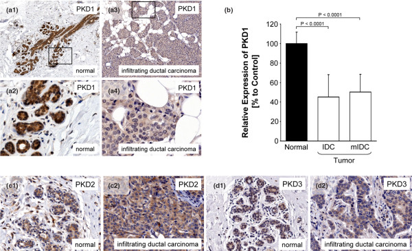
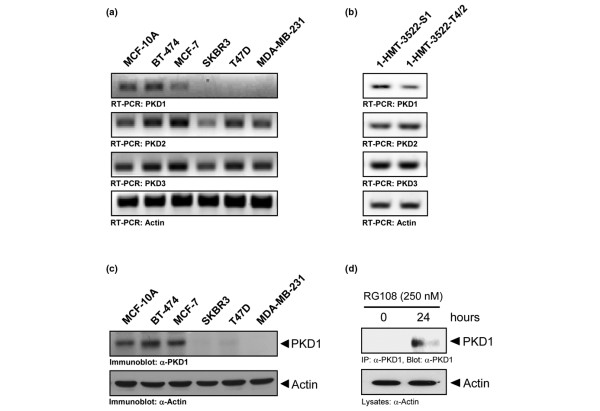
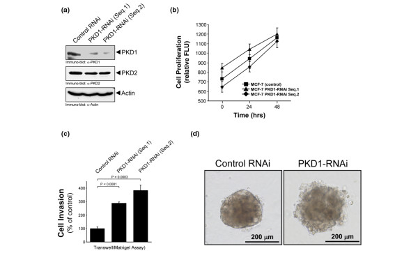
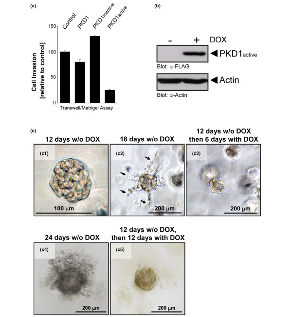
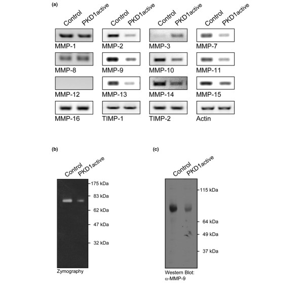
Results: We found that the serine/threonine kinase, PKD1, is highly expressed in ductal epithelial cells of normal human breast tissue, but is reduced in its expression in more than 95% of all analysed samples of human invasive breast tumours. Additionally, PKD1 is not expressed in highly invasive breast cancer cell lines, whereas non-invasive or very low-invasive breast cancer cell lines express PKD1. Our results further implicate that in MDA-MB-231 cells PKD1 expression is blocked by epigenetic silencing via DNA methylation. The re-expression of constitutively-active PKD1 in MDA-MB-231 cells drastically reduced their ability to invade in 2D and 3D cell culture. Moreover, MCF-7 cells acquired the ability to invade in 2D and 3D cell culture when PKD1 expression was knocked-down by shRNA. PKD1 also regulated the expression of breast cancer cell MMPs, MMP-2, MMP-7, MMP-9, MMP-10, MMP-11, MMP-13, MMP-14 and MMP-15, providing a potential mechanism for PKD1 mediation of the invasive phenotype.

Conclusions: Our results identify decreased expression of the PKD1 as a marker for invasive breast cancer. They further suggest that the loss of PKD1 expression increases the malignant potential of breast cancer cells. This may be due to the function of PKD1 as a negative regulator of MMP expression. Our data suggest re-expression of PKD1 as a potential therapeutic strategy.


Figures

Figure 1
Figure 1
PKD1 expression is reduced in invasive ductal carcinoma. (a to d) Tissue microarrays (TMAs) including 10 normal breast tissue samples, 40 invasive ductal carcinoma of the breast and 10 metastatic invasive ductal carcinoma samples from lymph nodes were analysed for the expression of the protein kinase D (PKD) family kinases PKD1, PKD2 and PKD3, using isoform specific antibodies. Representative pictures of normal and tumour tissue are depicted. (b) Statistical analysis of the normal and tumour samples. Error bars shown represent standard deviation. P values were acquired with the student's t-test using Graph Pad software. P values indicate extreme statistical significance. IDC = invasive ductal carcinoma; mIDC = metastatic invasive ductal carcinoma.
Figure 2
Figure 2
PKD1 is not expressed in invasive breast cancer cell lines. (a,b) MCF-10A cells and BT-474, MCF-7, SKBR3, T47D, MDA-MB-231, 1-HMT-3522-S1 or 1-HMT-3522-T4/2 breast cancer cell lines were cultivated under normal growth conditions. mRNA was isolated and the expression of protein kinase D (PKD) 1, PKD2, PKD3 and actin was detected by RT-PCR. (c) MCF-10A cells and BT-474, MCF-7, SKBR3, T47D, MDA-MB-231, 1-HMT-3522-S1 or 1-HMT-3522-T4/2 breast cancer cell lines were cultivated under normal growth conditions. Cells were lysed and lysates were analysed for PKD1 expression by western blotting. Actin served as loading control. (d) MDA-MB-231 cells were either left untreated or treated with RG108 (250 nM, for 24 hours). PKD1 was immunoprecipitated (α-PKD1 antibody) and immunoprecipitates were analysed for PKD1 expression. Western blotting of lysates for actin (α-actin antibody) served as a control.
Figure 3
Figure 3
The knockdown of PKD1 increases cell invasion. (a) MCF-7 cells were stably transduced with lentivirus coding for two different human shRNA sequences for protein kinase D (PKD) 1 (PKD1-RNAi Seq.1 and Seq.2) or for a non-target sequence which served as control. Cells were lysed and analysed for PKD1 expression by western blotting. Immunostaining for PKD2 and actin expression served as controls. (b) MCF-7 control and PKD1-RNAi clones were subjected to a cell proliferation assay using the CyQuant cell proliferation assay kit. (c) MCF-7 control and PKD1-RNAi clones were seeded on Matrigel-coated Transwell filters and Transwell invasion assays were performed over a time period of 16 hours. (d) MCF-7 cells stably-transfected with control RNAi or PKD1-RNAi were grown in 3D/Matrigel cell culture over a period of 60 days. Cells were photographed at day 60. Bars indicate the size of the spheroids. For experiments shown in b and c, error bars represent standard deviation. P values were acquired with the student's t-test using Graph Pad software. All P values indicate statistical significance.
Figure 4
Figure 4
Active PKD1 inhibits breast tumour cell invasion. (a) MDA-MB-231 cells were transiently transfected with wildtype protein kinase D (PKD) 1, kinase-dead PKD1 (PKD1inactive) or constitutively-active PKD1 (PKD1active). After 24 hours cells were seeded on Matrigel-coated Transwell filters and Transwell invasion assays were performed over a time period of 16 hours. (b) Inducible expression of active PKD1 in MDA-MB-231-TR-PKD1active (PKD1.Y463E mutant) cells. Cells were treated with doxycyclin (DOX) for 16 hours. Cells were lysed and lysates were analysed by western blotting for expression of constitutively-active PKD1 (anti-FLAG) or actin (loading control). (c) MDA-MB-231-TR-PKD1.Y463E cells were seeded in 3D culture and were either left untreated for 12 (c1), 18 (c2) and 24 days (c4), or were treated with doxycyclin after 12 days of normal growth to induce the expression of active PKD1 (c3 and c5). Cells were photographed at day 12 (c1), day 18 (c2 and c3) or day 24 (c4 and c5). Arrows in c2 indicate cells invading from the spheroid into the extracellular matrix. Bars indicate the size of the spheroids.
Figure 5
Figure 5
Active PKD1 regulates the expression of invasion-relevant MMPs. (a) MDA-MB-231 cells were transfected with vector control or constitutively-active protein kinase D (PKD) 1 (PKD1active, PKD1.Y463E mutant). After 16 hours mRNA was isolated and the expression of the matrix mettaloproteinases (MMP) MMP-1, MMP-2, MMP-3, MMP-7, MMP-8, MMP-9, MMP-10, MMP-11, MMP-12, MMP-13, MMP-14, MMP-15 and MMP-16, tissue inhibitors of metalloproteinases (TIMP) TIMP1 and TIMP2 and actin (control) was analysed by RT-PCR (shown as a PCR reaction with 35 cycles; PCR reaction with 20 and 40 cycles showed similar results). (b,c) MDA-MB-231 cells were transfected with vector control or constitutively-active PKD1 (PKD1active, PKD1.Y463E mutant). After 48 hours supernatants were collected and used for (b) zymographic analysis or (c) western blot analysis for MMP-9 expression.

References

    1. Storz P, Doppler H, Toker A. Protein kinase D mediates mitochondrion-to-nucleus signaling and detoxification from mitochondrial reactive oxygen species. Mol Cell Biol. 2005;25:8520–8530. doi: 10.1128/MCB.25.19.8520-8530.2005. - DOI - PMC - PubMed
    1. Rykx A, De Kimpe L, Mikhalap S, Vantus T, Seufferlein T, Vandenheede JR, Van Lint J. Protein kinase D: a family affair. FEBS letters. 2003;546:81–86. doi: 10.1016/S0014-5793(03)00487-3. - DOI - PubMed
    1. Hausser A, Storz P, Martens S, Link G, Toker A, Pfizenmaier K. Protein kinase D regulates vesicular transport by phosphorylating and activating phosphatidylinositol-4 kinase IIIbeta at the Golgi complex. Nat Cell Biol. 2005;7:880–886. doi: 10.1038/ncb1289. - DOI - PMC - PubMed
    1. Bowden ET, Barth M, Thomas D, Glazer RI, Mueller SC. An invasion-related complex of cortactin, paxillin and PKCmu associates with invadopodia at sites of extracellular matrix degradation. Oncogene. 1999;18:4440–4449. doi: 10.1038/sj.onc.1202827. - DOI - PubMed
    1. Endo K, Oki E, Biedermann V, Kojima H, Yoshida K, Johannes FJ, Kufe D, Datta R. Proteolytic cleavage and activation of protein kinase C μ by caspase-3 in the apoptotic response of cells to 1-beta-D-arabinofuranosylcytosine and other genotoxic agents. J Biol Chem. 2000;275:18476–18481. doi: 10.1074/jbc.M002266200. - DOI - PubMed

Publication types

MeSH terms