. 2009 Feb 25:7:19.
doi: 10.1186/1477-7827-7-19.

Structural alterations in the seminiferous tubules of rats treated with immunosuppressor tacrolimus

Affiliations

Structural alterations in the seminiferous tubules of rats treated with immunosuppressor tacrolimus

Breno H Caneguim et al. Reprod Biol Endocrinol. .

Abstract

Background: Tacrolimus (FK-506) is an immunosuppressant that binds to a specific immunophilin, resulting in the suppression of the cellular immune response during transplant rejection. Except for some alterations in the spermatozoa, testicular morphological alterations have not been described in rats treated with tacrolimus. In the present study, we purpose to evaluate if the treatment with tacrolimus at long term of follow-up interferes in the integrity of the seminiferous tubules.

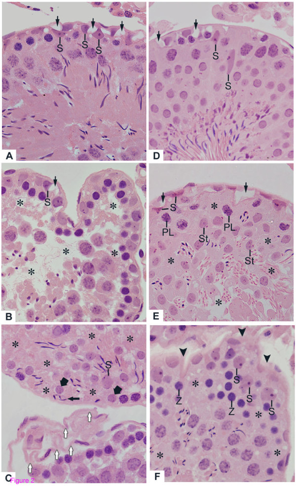
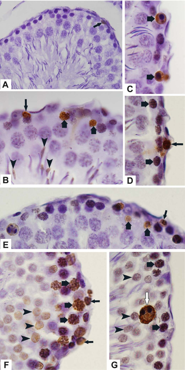
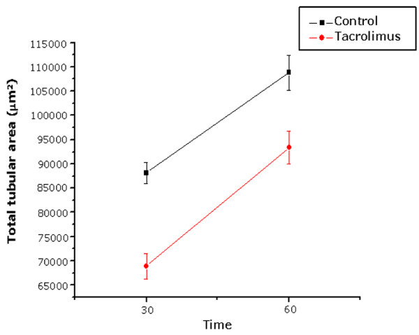
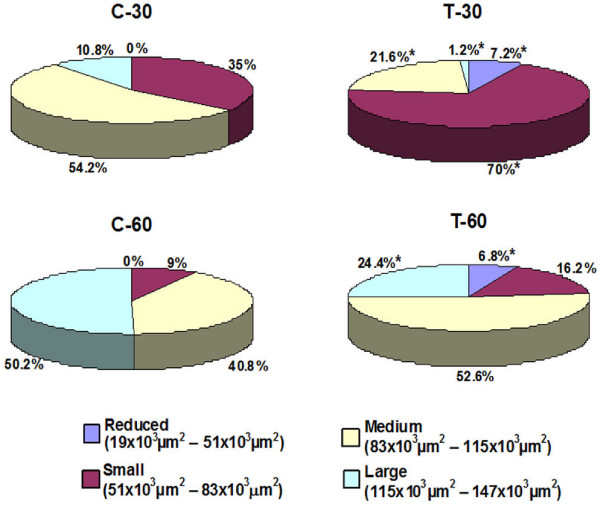
Methods: Rats aging 42-day-old received daily subcutaneous injections of 1 mg/kg/day of tacrolimus during 30 (T-30) and 60 (T-60) days; the rats from control groups (C-30 and C-60) received saline solution. The left testes were fixed in 4% formaldehyde and embedded in glycol methacrylate for morphological and morphometric analyses while right testes were fixed in Bouin's liquid and embedded in paraffin for detection of cell death by the TUNEL method. The epithelial and total tubular areas as well as the stages of the seminiferous epithelium and the number of spermatocytes, spermatids and Sertoli cells (SC) per tubule were obtained.

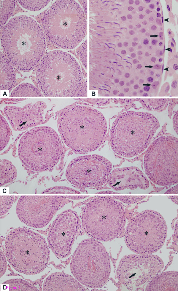
Results: In the treated groups, seminiferous tubules irregularly outlined showed disarranged cellular layers and loss of germ cells probably due to cell death, which was revealed by TUNEL method. In addition to germ cells, structural alterations in the SC and folding of the peritubular tissue were usually observed. The morphometric results revealed significant decrease in the number of SC, spermatocytes, spermatids and significant reduction in the epithelial and total tubular areas.

Conclusion: Tacrolimus induces significant histopathological disorders in the seminiferous tubules, resulting in spermatogenic damage and reduction in the number of Sertoli cells. A careful evaluation of the peritubular components will be necessary to clarify if these alterations are related to the effect of FK-506 on the peritubular tissue.


Figures

Figure 1
Figure 1
Photomicrographs of seminiferous tubules of rats from C-30 (A), C-60 (B), T-30 (C) and T-60 (D) groups stained by H.E. (A and B) In the seminiferous tubules with normal aspect, the germ cells are organized in concentric layers and the tubular lumen is empty (A, asterisks); (B) The Sertoli cell nuclei (arrows) are positioned adjacent to the well defined peritubular tissue in which peritubular cells are observed (arrowheads). In T-30 (C) and T-60 (D), the altered seminiferous tubules show irregular shape, epithelial disorganization and detached germ cells filling the tubular lumen (asterisks). In some atrophied seminiferous tubules, loss of germ cells is observed (arrows). Figs. 1A, 1C and 1D: ×110; Figs. 1B: ×330.
Figure 2
Figure 2
Photomicrographs of seminiferous tubules of rats from T-30 (A, B and C) and T-60 (D, E and F) groups stained by H.E. (A and B) Slightly (A) and severely (B) damaged tubules show vacuolar spaces (arrows) adjacent to Sertoli cell nuclei with irregular shape (S). In B, intraepithelial spaces (asterisks) due to lack of spermatocytes and spermatids were observed. (C) Lack of germ cells (asterisks) is noted in the basal and adluminal portions. Round (thick arrows) and elongate (thin arrow) spermatids are abnormally positioned in the basal compartment. Note a single Sertoli cell showing dislocated nucleus (S). In the other tubule, the peritubular tissue is intensely folded (white arrows). (D) Slightly altered seminiferous tubule shows vacuolar spaces (arrows) adjacent to Sertoli cell nuclei. Some irregular Sertoli cell nuclei (S) are displaced from their original site. (E and F) The damaged tubules show lack of germ cells (asterisks) in the layers of round spermatids (St, in E), pre-leptotene (PL, in E) and zygotene (Z, in F) spermatocytes. In E, vacuolar spaces (arrows) adjacent to irregular and strongly stained Sertoli cell nuclei (S) are observed. In F, the peritubular tissue is irregularly outlined (arrowheads). Adjacent to this altered tissue, the Sertoli cell nuclei are irregular and displaced from their original site (S). Figs. 2A-2F: ×330.
Figure 3
Figure 3
Photomicrographs of seminiferous tubules of rats from C-30 (A), T-30 (B-D) and T-60 (E-G) submitted to the TUNEL-method. (A) TUNEL-positive germ cell is observed in the epithelium (arrow). (B-D) Primary spermatocytes (thick arrows), spermatogonia (thin arrows) and elongate spermatids (arrowheads) are labeled by TUNEL method. (E-G) Spermatogonia (thin arrows), primary spermatocytes in different stages (thick arrows) and round spermatids (arrowheads) are TUNEL-positive. A giant multinucleated cell derivative from round spermatids (white arrow) is also positive. Fig. 3A: ×260; Figs. 3B-3G: ×710.
Figure 4
Figure 4
Effect of FK-506 upon total tubular area (μm2) of the seminiferous tubules according to the time of treatment.
Figure 5
Figure 5
Frequency (%) of seminiferous tubules according to the total tubular area (μm2) of animals from control (C-30 and C-60) and tacrolimus (T-30 and T-60) groups. *p < 0.05 (statistically significant).

References

    1. Plosker GL, Foster RH. Tacrolimus: a further update of its pharmacology and therapeutic use in the management of organ transplantation. Drugs. 2000;59:323–389. doi: 10.2165/00003495-200059020-00021. - DOI - PubMed
    1. Kino T, Hatanaka H, Hashimoto M, Nishiyama M, Goto T, Okuhara M, Kohsaka M, Aoki H, Imanaka H. FK-506, a novel immunosuppressant isolated from a Streptomyces. I. Fermentation, isolation, and physico-chemical and biological characteristics. J Antibiot. 1987;40:1249–1255. - PubMed
    1. Christians U, Jacobsen W, Benet LZ, Lampen A. Mechanisms of clinically relevant drug interactions associated with tacrolimus. Clin Pharmacokinet. 2002;41:813–851. doi: 10.2165/00003088-200241110-00003. - DOI - PubMed
    1. Venkataramanan R, Swaminathan A, Prasad T, Jain A, Zuckerman S, Warty V, McMichael J, Lever J, Burckart G, Starzl T. Clinical pharmacokinetics of Tacrolimus. Clin Pharmacokinet. 1995;29:404–430. doi: 10.2165/00003088-199529060-00003. - DOI - PubMed
    1. Nagase K, Iwasaki K, Nozaki K, Noda K. Distribution and protein binding of FK506, a potent immunosuppressive macrolide lactone, in human blood and its uptake by erythrocytes. J Pharm Pharmacol. 1994;46:113–117. - PubMed

Publication types

LinkOut - more resources