. 2009 Nov;41(5):562-72.
doi: 10.1165/rcmb.2008-0377OC. Epub 2009 Feb 24.

Rapamycin prevents transforming growth factor-alpha-induced pulmonary fibrosis

Affiliations

Rapamycin prevents transforming growth factor-alpha-induced pulmonary fibrosis

Thomas R Korfhagen et al. Am J Respir Cell Mol Biol. 2009 Nov.

Abstract

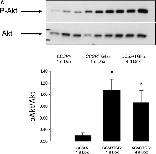
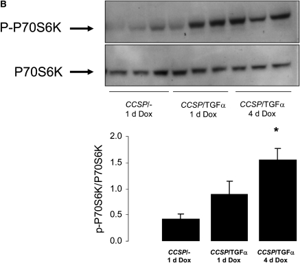
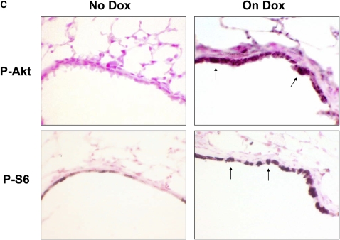
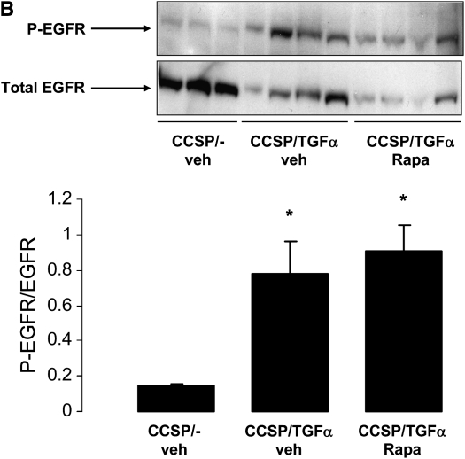
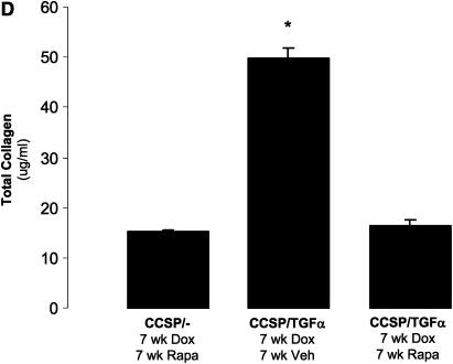
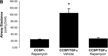
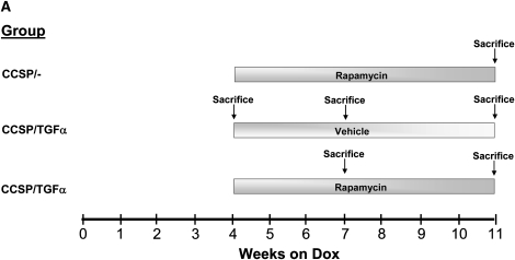
Transforming growth factor (TGF)-alpha is a ligand for the epidermal growth factor receptor (EGFR). EGFR activation is associated with fibroproliferative processes in human lung disease and animal models of pulmonary fibrosis. Overexpression of TGF-alpha in transgenic mice causes progressive and severe pulmonary fibrosis; however, the intracellular signaling pathways downstream of EGFR mediating this response are unknown. Using a doxycycline-regulatable transgenic mouse model of lung-specific TGF-alpha expression, we observed increased PCNA protein and phosphorylation of Akt and p70S6K in whole lung homogenates in association with induction of TGF-alpha. Induction in the lung of TGF-alpha caused progressive pulmonary fibrosis over a 7-week period. Daily administration of rapamycin prevented accumulation of total lung collagen, weight loss, and changes in pulmonary mechanics. Treatment of mice with rapamycin 4 weeks after the induction of TGF-alpha prevented additional weight loss, increases in total collagen, and changes in pulmonary mechanics. Rapamycin prevented further increases in established pulmonary fibrosis induced by EGFR activation. This study demonstrates that mammalian target of rapamycin (mTOR) is a major effector of EGFR-induced pulmonary fibrosis, providing support for further studies to determine the role of mTOR in the pathogenesis and treatment of pulmonary fibrosis.


Figures

Figure 1.
Figure 1.
Transforming growth factor (TGF)-α induces phosphorylation of the Akt, P70S6K, and S6. Epithelial cell TGF-α expression increased phosphatidylinositol 3-kinase (PI3K)/mammalian target of rapamycin (mTOR) activity as assessed by increased phosphorylation of Akt, P70S6K, and S6. CCSP/TGF-α mice were treated with doxycycline (Dox) to induce TGF-α expression for 1 or 4 days and compared with littermate control single transgene CCSP/- mice treated with 1 day of Dox. Immunoblotting of lung homogenates from CCSP/TGF-α mice demonstrated (A) increased phosphorylation of Akt at Ser473 and (B) phosphorylation of P70S6K at Thr389. Immunostaining for phosphorylated Akt and phosphorylated S6 demonstrated increased staining in the airway epithelium in CCSP/TGF-α mice after 4 days of Dox compared with CCSP/TGF-α mice not receiving Dox (C, arrows). Mean ± SE, n = 3–6 mice of each genotype; * P < 0.05.
Figure 1.
Figure 1.
Transforming growth factor (TGF)-α induces phosphorylation of the Akt, P70S6K, and S6. Epithelial cell TGF-α expression increased phosphatidylinositol 3-kinase (PI3K)/mammalian target of rapamycin (mTOR) activity as assessed by increased phosphorylation of Akt, P70S6K, and S6. CCSP/TGF-α mice were treated with doxycycline (Dox) to induce TGF-α expression for 1 or 4 days and compared with littermate control single transgene CCSP/- mice treated with 1 day of Dox. Immunoblotting of lung homogenates from CCSP/TGF-α mice demonstrated (A) increased phosphorylation of Akt at Ser473 and (B) phosphorylation of P70S6K at Thr389. Immunostaining for phosphorylated Akt and phosphorylated S6 demonstrated increased staining in the airway epithelium in CCSP/TGF-α mice after 4 days of Dox compared with CCSP/TGF-α mice not receiving Dox (C, arrows). Mean ± SE, n = 3–6 mice of each genotype; * P < 0.05.
Figure 2.
Figure 2.
Erlotinib and rapamycin inhibit TGF-α–induced phosphorylation of P70S6K and PCNA. (A) Pretreatment of CCSP/TGF-α mice with erlotinib (100 mg/kg) prevented increased phosphorylation of Akt and P70S6K and increased levels of PCNA after 1 day of Dox. Pretreatment of CCSP/TGF-α mice with rapamycin (Rapa, 4 mg/kg) did not alter phosphorylation of Akt, but prevented increased phosphorylation of P70S6K and increased levels of PCNA after 1 day of Dox. (B) Rapamycin did not effect phosphorylation of EGFR (Y1086) in lung homogenates after 1 day of Dox. Mean ± SE, *P < 0.05 compared with vehicle (veh)-treated (the rapamycin vehicle or 0.25% PEG400, 0.25% Tween 20) CCSP/- controls.
Figure 2.
Figure 2.
Erlotinib and rapamycin inhibit TGF-α–induced phosphorylation of P70S6K and PCNA. (A) Pretreatment of CCSP/TGF-α mice with erlotinib (100 mg/kg) prevented increased phosphorylation of Akt and P70S6K and increased levels of PCNA after 1 day of Dox. Pretreatment of CCSP/TGF-α mice with rapamycin (Rapa, 4 mg/kg) did not alter phosphorylation of Akt, but prevented increased phosphorylation of P70S6K and increased levels of PCNA after 1 day of Dox. (B) Rapamycin did not effect phosphorylation of EGFR (Y1086) in lung homogenates after 1 day of Dox. Mean ± SE, *P < 0.05 compared with vehicle (veh)-treated (the rapamycin vehicle or 0.25% PEG400, 0.25% Tween 20) CCSP/- controls.
Figure 3.
Figure 3.
Rapamycin prevents TGF-α–dependent pulmonary fibrosis. CCSP/TGF-α mice were administered 7 weeks of Dox, and treated with either rapamycin (4 mg/kg once daily 6 d/wk) or vehicle. Controls were littermate single transgene CCSP/- mice administered Dox and treated with rapamycin. The treatment protocol is represented schematically in A. (B) Rapamycin prevented TGF-α–induced decreases in progressive body weight loss. Sections of lungs were stained with hematoxylin and eosin. (C) CCSP/TGF-α mice administered rapamycin at the initiation of Dox caused a marked attenuation of pulmonary fibrosis compared with CCSP/TGF-α vehicle-treated mice. (D) Rapamycin prevented TGF-α–induced increases in total lung collagen. Photomicrographs shown are representative of lungs from seven mice in each group. All photomicrographs are taken at the same magnification, and bar is 200 μm. *P < 0.05 compared with CCSP/- control and CCSP/TGF-α vehicle-treated mice.
Figure 3.
Figure 3.
Rapamycin prevents TGF-α–dependent pulmonary fibrosis. CCSP/TGF-α mice were administered 7 weeks of Dox, and treated with either rapamycin (4 mg/kg once daily 6 d/wk) or vehicle. Controls were littermate single transgene CCSP/- mice administered Dox and treated with rapamycin. The treatment protocol is represented schematically in A. (B) Rapamycin prevented TGF-α–induced decreases in progressive body weight loss. Sections of lungs were stained with hematoxylin and eosin. (C) CCSP/TGF-α mice administered rapamycin at the initiation of Dox caused a marked attenuation of pulmonary fibrosis compared with CCSP/TGF-α vehicle-treated mice. (D) Rapamycin prevented TGF-α–induced increases in total lung collagen. Photomicrographs shown are representative of lungs from seven mice in each group. All photomicrographs are taken at the same magnification, and bar is 200 μm. *P < 0.05 compared with CCSP/- control and CCSP/TGF-α vehicle-treated mice.
Figure 4.
Figure 4.
Rapamycin prevents TGF-α–dependent changes in lung mechanics. Rapamycin administered daily to CCSP/TGF-α mice at the time of Dox treatment prevented TGF-α–mediated (A–C) increases in airway resistance, and airway and tissue elastance, and (D) decreases in compliance compared with CCSP/TGF-α receiving vehicle. *P < 0.05 compared with CCSP/- control and CCSP/TGF-α vehicle-treated mice.
Figure 4.
Figure 4.
Rapamycin prevents TGF-α–dependent changes in lung mechanics. Rapamycin administered daily to CCSP/TGF-α mice at the time of Dox treatment prevented TGF-α–mediated (A–C) increases in airway resistance, and airway and tissue elastance, and (D) decreases in compliance compared with CCSP/TGF-α receiving vehicle. *P < 0.05 compared with CCSP/- control and CCSP/TGF-α vehicle-treated mice.
Figure 5.
Figure 5.
Rapamycin prevents progression of TGF-α–dependent pulmonary fibrosis. After 4 weeks of Dox, CCSP/TGF-α mice were administered either daily rapamycin or vehicle. Rapamycin-treated mice were evaluated after 3 or 7 weeks of treatment. Vehicle-treated CCSP/TGF-α mice were recovered at 4, 7, and 11 weeks of Dox for comparison. Control mice were CCSP/- mice treated with rapamycin after 4 weeks of Dox. The treatment protocol is represented schematically in A. (B) Dox-induced expression of TGF-α caused progressive weight loss. Body weight stabilized in mice treated with rapamycin 4 weeks after treatment with Dox. Sections of lungs were stained with hematoxylin and eosin. (C) Expression of TGF-α caused progressive adventitial fibrosis. CCSP/TGF-α mice administered rapamycin after 4 weeks of Dox demonstrated reduced (C) adventitial fibrosis and (D) total lung collagen compared with CCSP/TGF-α vehicle-treated mice receiving 7 or 11 weeks of Dox. *P < 0.05 compared with rapamycin-treated controls. +P < 0.05 compared with 7 and 11 week vehicle-treated CCSP/TGF-α mice. Small images of dead mice in B denote mouse death during week.
Figure 5.
Figure 5.
Rapamycin prevents progression of TGF-α–dependent pulmonary fibrosis. After 4 weeks of Dox, CCSP/TGF-α mice were administered either daily rapamycin or vehicle. Rapamycin-treated mice were evaluated after 3 or 7 weeks of treatment. Vehicle-treated CCSP/TGF-α mice were recovered at 4, 7, and 11 weeks of Dox for comparison. Control mice were CCSP/- mice treated with rapamycin after 4 weeks of Dox. The treatment protocol is represented schematically in A. (B) Dox-induced expression of TGF-α caused progressive weight loss. Body weight stabilized in mice treated with rapamycin 4 weeks after treatment with Dox. Sections of lungs were stained with hematoxylin and eosin. (C) Expression of TGF-α caused progressive adventitial fibrosis. CCSP/TGF-α mice administered rapamycin after 4 weeks of Dox demonstrated reduced (C) adventitial fibrosis and (D) total lung collagen compared with CCSP/TGF-α vehicle-treated mice receiving 7 or 11 weeks of Dox. *P < 0.05 compared with rapamycin-treated controls. +P < 0.05 compared with 7 and 11 week vehicle-treated CCSP/TGF-α mice. Small images of dead mice in B denote mouse death during week.
Figure 6.
Figure 6.
Rapamycin prevents progression of TGF-α–dependent changes in lung mechanics. CCSP/TGF-α transgenic mice administered rapamycin 4 weeks after treatment with Dox demonstrated (A–C) reduced increases in airway resistance, and airway and tissue elastance, and (D) decreases in compliance compared with vehicle-treated CCSP/TGF-α mice receiving 7 and 11 weeks of Dox. *P < 0.05 compared with CCSP/- control mice; + P < 0.05 compared with 7 and 11 week vehicle-treated CCSP/TGF-α transgenic mice.
Figure 6.
Figure 6.
Rapamycin prevents progression of TGF-α–dependent changes in lung mechanics. CCSP/TGF-α transgenic mice administered rapamycin 4 weeks after treatment with Dox demonstrated (A–C) reduced increases in airway resistance, and airway and tissue elastance, and (D) decreases in compliance compared with vehicle-treated CCSP/TGF-α mice receiving 7 and 11 weeks of Dox. *P < 0.05 compared with CCSP/- control mice; + P < 0.05 compared with 7 and 11 week vehicle-treated CCSP/TGF-α transgenic mice.

References

    1. Cook DN, Brass DM, Schwartz DA. A matrix for new ideas in pulmonary fibrosis. Am J Respir Cell Mol Biol 2002;27:122–124. - PubMed
    1. Crystal RG, Gadek JE, Ferrans VJ, Fulmer JD, Line BR, Hunninghake GW. Interstitial lung disease: current concepts of pathogenesis, staging and therapy. Am J Med 1981;70:542–568. - PubMed
    1. Crystal RG, Bitterman PB, Mossman B, Schwarz MI, Sheppard D, Almasy L, Chapman HA, Friedman SL, King TE Jr, Leinwand LA, et al. Future research directions in idiopathic pulmonary fibrosis: summary of a National Heart, Lung, and Blood Institute working group. Am J Respir Crit Care Med 2002;166:236–246. - PubMed
    1. Kuwano K, Hagimoto N, Hara N. Molecular mechanisms of pulmonary fibrosis and current treatment. Curr Mol Med 2001;1:551–573. - PubMed
    1. Hardie WD, Le Cras TD, Jiang K, Tichelaar JW, Azhar M, Korfhagen TR. Conditional expression of transforming growth factor-alpha in adult mouse lung causes pulmonary fibrosis. Am J Physiol Lung Cell Mol Physiol 2004;286:L741–L749. - PubMed

Publication types

MeSH terms

Substances