. 2009 Apr 17;284(16):10968-79.
doi: 10.1074/jbc.M804923200. Epub 2009 Feb 25.

Wnt-5a-CKI{alpha} signaling promotes {beta}-catenin/E-cadherin complex formation and intercellular adhesion in human breast epithelial cells

Affiliations

Wnt-5a-CKI{alpha} signaling promotes {beta}-catenin/E-cadherin complex formation and intercellular adhesion in human breast epithelial cells

Catharina Medrek et al. J Biol Chem. .

Abstract

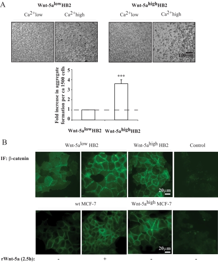
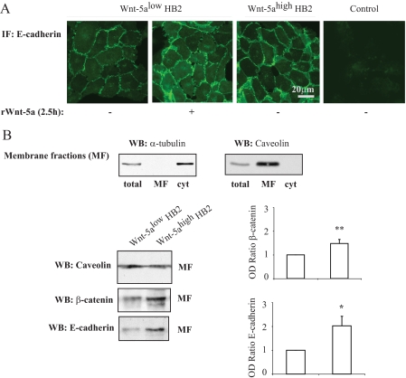
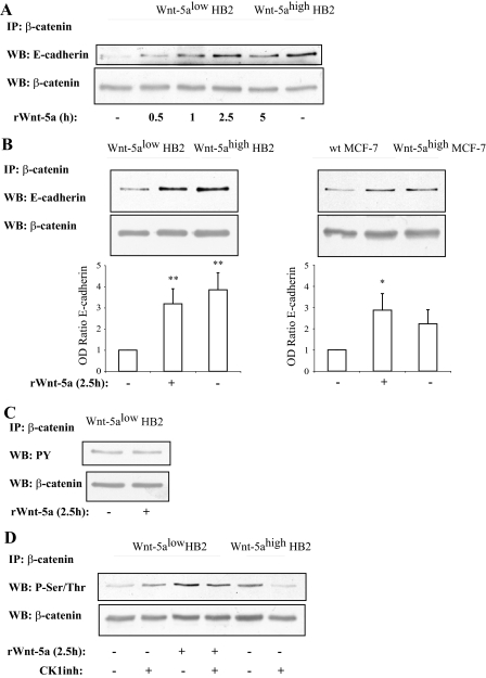
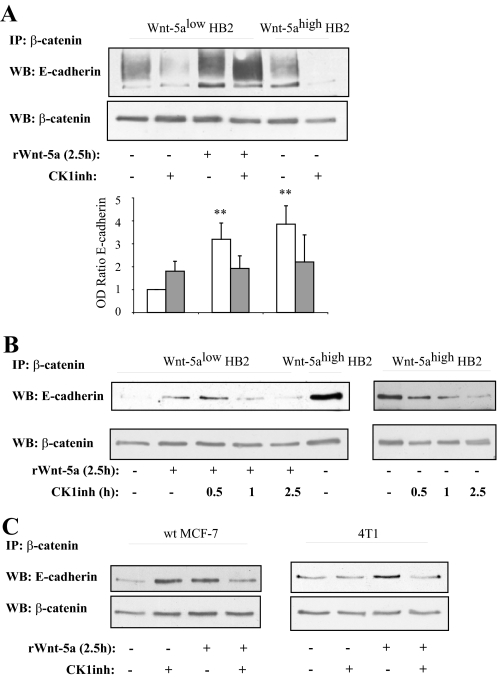
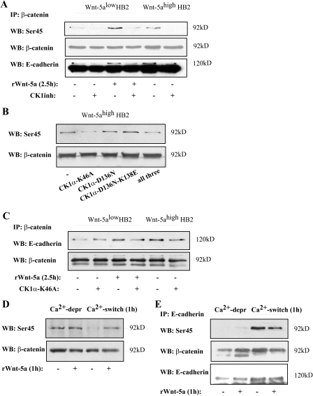
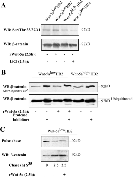
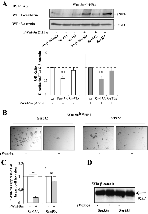
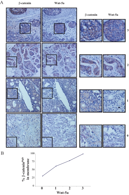
Wnt-5a is a non-transforming Wnt protein that is implicated in cell polarity, adhesion, and motility. We have previously shown that low expression of Wnt-5a is a predictor of shorter disease-free survival in human breast cancer. Here, we investigated whether beta-catenin/E-cadherin-mediated cell-cell adhesion was affected by loss of Wnt-5a in breast carcinomas, thereby promoting a metastatic behavior of the tumor. We show that Wnt-5a stimulation of human breast epithelial cells leads to an increased Ca(2+)-dependent cell-cell adhesion. Furthermore, Wnt-5a/casein kinase Ialpha (CKIalpha)-specific Ser-45 phosphorylation of beta-catenin is associated with an increased complex formation of beta-catenin/E-cadherin. Mutation of Ser-45 decreases the beta-catenin/E-cadherin association. Also, the inhibitory effect of Wnt-5a on breast epithelial cell invasion is reduced upon mutation of beta-catenin-Ser-45. The Wnt-5a-CKIalpha-induced Ser-45 phosphorylation does not lead to degradation of beta-catenin. Finally we show that human breast cancers lacking Wnt-5a protein have a significantly lower level of membrane-associated beta-catenin. Down-regulation of Wnt-5a expression and subsequent reduction of membrane-associated beta-catenin in invasive breast cancer, can therefore contribute to a decreased cell-cell adhesion and increased motility resulting in a higher probability for metastatic disease.


Figures

FIGURE 1.
FIGURE 1.
A, cell aggregation assay. Human breast epithelial cells with varying Wnt-5a protein expression levels of HB2 Wnt-5alow and HB2 Wnt-5ahigh cells were treated with Versene, resuspended in Ca2+ high as compared with Ca2+ low medium and allowed to form aggregates for 1 h upon slow rocking. HB2 Wnt-5ahigh cells formed larger cell-cell clusters than HB2 Wnt-5alow cells. The levels of propidium iodide staining were equal. The diagram represents the fold-increase in number of aggregates formed per approximately 1500 cells in HB2 Wnt-5ahigh compared with HB2 Wnt-5alow cells. Error bars indicate S.E., n = 7, ***, p < 0.001. B, immunofluorescence (IF) studies of β-catenin membrane localization in human breast epithelial cells with varying Wnt-5a protein expression levels or treated with recombinant Wnt-5a. HB2 Wnt-5alow, HB2 Wnt-5alow treated with recombinant Wnt-5a and HB2 Wnt-5ahigh (upper); MCF-7, MCF-7 and treated with recombinant Wnt-5a and MCF-7 Wnt-5ahigh (lower)(n = 5).
FIGURE 2.
FIGURE 2.
A, immunofluorescence (IF) studies of E-cadherin membrane localization in human breast epithelial cells (HB2) with varying Wnt-5a protein expression levels or in HB2 Wnt-5alow cells treated with recombinant Wnt-5a (rWnt-5a)(n = 3). B, membrane fractionation experiments. The purity of the membrane fractions (total cell lysate (total), membrane fraction (MF), and cytosol (cyt)) was analyzed with α-tubulin and caveolin antibodies (upper panel). Western blot analysis of E-cadherin and β-catenin protein expression levels in membrane fractions from human breast epithelial cells with varying Wnt-5a protein expression levels (lower panels). To ensure equal loading, protein measurements were done using a Coomassie® protein assay reagent (Pierce). The blots shown are representative of several separate experiments and OD measurements of the band intensities were performed to quantify the differences (right panel, histograms). Error bars indicate S.E., n = 16; *, p < 0.05; **, p < 0.01. WB, Western blot.
FIGURE 3.
FIGURE 3.
A, time kinetics of Wnt-5a inducedβ-catenin/E-cadherin complex formation indicates that it is a rapid and specific process unrelated to general protein expression levels (n = 3). B, β-catenin/E-cadherin complex formation is potentiated by Wnt-5a signaling as judged by co-immunoprecipitations of β-catenin/E-cadherin complexes from human mammary epithelial cell line HB2 (left) and human breast cancer cell line MCF-7 (right) cells with varying Wnt-5a expression levels or in cells treated with recombinant Wnt-5a. The blot shown is representative of several separate experiments and OD measurements of the band intensities were performed to quantify the differences (lower panel; histograms). Error bars indicate S.E., n = 8, n = 5; *, p < 0.05; **, p < 0.01. C, tyrosine phosphorylation of β-catenin is not affected by Wnt-5a stimulation, whereas serine/threonine phosphorylation is (D) as judged by immunoprecipitation of β-catenin and Western blot (WB) using a total Tyr(P) or Ser/Thr antibodies. Inhibition of the serine/threonine kinase CKI, with the casein kinase 1 α/ε inhibitor IC261, disrupts the Wnt-5a-induced Ser/Thr phosphorylation of β-catenin, whereas cells lacking Wnt-5a remain Ser/Thr phosphorylated on β-catenin (D). The CKI inhibitor was added for 1 h at the end of the incubation with rWnt-5a (n = 5). Wt, wild type.
FIGURE 4.
FIGURE 4.
A, the Wnt-5a-induced β-catenin/E-cadherin complex formation is specifically inhibited by the CKI inhibitor only in cells treated with, or expressing, Wnt-5a. The blot shown is representative of several separate experiments and OD measurements of the band intensities were performed to quantify the differences (lower panel, histograms). Error bars indicate S.E., n = 8; *, p < 0.05; **, p < 0.01. B, time kinetics of treatment with the CKI inhibitor indicates that Wnt-5a is upstream of CKI in induction of β-catenin/E-cadherin complex formation (n = 3). C, the Wnt-5a/CKI induced β-catenin/E-cadherin complex formation is also seen in two different breast cancer cell lines: MCF-7 (left) and 4T1 (right). The blot shown is representative of several separate experiments and OD measurements of the band intensities were performed to quantify the differences (supplemental Fig. S2A). WB, Western blot; IP, immunoprecipitate.
FIGURE 5.
FIGURE 5.
A, Wnt-5a/CKI specifically induces Ser-45 phosphorylation of β-catenin. This correlates with increased levels of co-immunoprecipitated E-cadherin (n = 3). The lower band in the E-cadherin reblot is β-catenin. B, transfection of Wnt-5ahigh HB2 cells with three different variants of dominant negative kinase-deficient mutants of CKIα (26): K46A, D136N, and D136N-K138E or all three. Overexpression of CKIα-K46A disrupts the Wnt-5a-induced Ser-45 phosphorylation of β-catenin. C, overexpression of CKIα-K46A also disrupts the Wnt-5a-induced increase in β-catenin/E-cadherin complex formation. D, initiation of cell-cell adhesion per se does not induce Ser-45 phosphorylation of β-catenin. Ca2+-dependent cell-cell adhesion was initiated by performing a Ca2+-switch experiment (see “Experimental Procedures”). Wnt-5alow HB2 cells were allowed to re-adhere for 1 h after the Ca2+ switch in the absence or presence of rWnt-5a. Cells where cell-cell contacts had recently been disrupted and cells treated with rWnt-5a show β-catenin Ser-45 phosphorylation (also see C). In sharp contrast, initiation of de novo cell-cell contacts per se does not lead to Ser-45 phosphorylation, whereas addition of rWnt-5a does (n = 3). E, the pool of Ser-45-phosphorylated β-catenin seen after cell-cell contact disruption in B is not associated to E-cadherin complexes as shown by co-immunoprecipitations of E-cadherin after Ca2+ deprivation. In sharp contrast, the E-cadherin-associated β-catenin after a Ca2+ switch (re-adherence) is Ser-45 phosphorylated. As described in the text it is not possible to compare the amount of coprecipitated β-catenin using the (SHE 78-7) E-cadherin ectodomain antibody and this might explain why we do not see an increased Ser-45 phosphorylation after Wnt-5a stimulation compared with unstimulated cells in this particular set of experiments. The lower band in the E-cadherin reblot is β-catenin. WB, Western blot; IP, immunoprecipitate.
FIGURE 6.
FIGURE 6.
A, Wnt-5a signaling does not induce Ser-33/Ser-37/Thr-41 phosphorylation in human breast epithelial cells. LiCl (10 μm) was used as a control. Note that the level of total β-catenin is slightly higher in the second lane of this particular experiment. B, analysis of β-catenin ubiquitination. HB2 Wnt-5alow cells and HB2 Wnt-5alow cells stimulated with rWnt-5a (2.5 h) or HB2 Wnt-5ahigh cells were treated with the protease inhibitor MG132 for 5 h prior to cell lysis (lysis buffer contained the protease inhibitor N-ethylmaleimide) and levels of ubiquitinated β-catenin were analyzed (n = 3). C, pulse-chase experiments were performed to analyze the rate of β-catenin degradation. Wnt-5a does not induce β-catenin degradation in human breast epithelial cells as compared with untreated cells. Longer chase periods gave similar results (data not shown). The level of total β-catenin was controlled by Western blot (WB) (lower panel).
FIGURE 7.
FIGURE 7.
A, co-immunoprecipitation of E-cadherin with different forms of FLAG-tagged β-catenin (35). HB2 Wnt-5alow cells were transfected with FLAG-tagged wild type (wt) β-catenin, Ser-45Δ-mutated β-catenin, and Ser-33Δ-mutated β-catenin. Immunoprecipitation was performed using anti-FLAG antibodies. Due to a decreased degradation of β-catenin harboring the Ser-45Δ and Ser-33Δ mutations, FLAG β-catenin had to be set to equal levels in the blots by a pre-Western blot (WB) analysis after which the level of coprecipitated E-cadherin compared with FLAG β-catenin in the final blot could be measured using OD measurements. The histogram thus shows the relative levels (OD E-cadherin/OD FLAG-β-catenin) of bound E-cadherin to FLAG-tagged wild type β-catenin as compared with FLAG-tagged Ser-45Δ and Ser-33Δ β-catenin, under normal (left) or Wnt-5a stimulating (right) conditions (***, p < 0.001 compared with wild type, S.E., n = 7). B, Matrigel-invasion assay. HB2 Wnt-5alow cells were transfected with FLAG-tagged Ser-33Δ β-catenin or Ser-45Δ β-catenin. After a 48-h transfection period the cells were detached, counted, and 25,000 cells/chamber were allowed to invade Matrigel invasion chambers in the absence or presence of rWnt-5a in the upper chamber. C, the histogram (representing B) shows the fold-reduction in number of invaded cells upon rWnt-5a treatment for both Ser-33Δ β-catenin- and Ser-45Δ β-catenin-transfected cells (**, p < 0.01; *, p < 0.05; S.E.; n = 3). D, the levels of FLAG-tagged β-catenin protein were similar between the samples in B and C.
FIGURE 8.
FIGURE 8.
A, summarizing presentation of immunostainings of Wnt-5a and membrane-associated β-catenin in representative sections of human invasive breast carcinomas. Staining was done with a polyclonal rabbit antibody against human Wnt-5a (19) and an antibody directed against β-catenin. The tumors in the figure represents gradings 0 (lower) to 3 (upper) (see “Experimental Procedures” for description) for membrane-associated β-catenin and Wnt-5a (left) with magnifications representing the squared fields (right). B, the diagram represents the percentage of β-cateninhigh tumors (grading 2 and 3) in the different Wnt-5a groups (0, 1, 2, 3). The statistical Chi-square test was used and the linear to linear association between Wnt-5a and membrane-bound β-catenin was p = 0.004.

References

    1. Osborne, M. P. (2000) Breast Anatomy and Development, 2nd Ed., Philadelphia, PA
    1. Berx, G., Nollet, F., and van Roy, F. (1998) Cell Adhes. Commun. 6 171-184 - PubMed
    1. Drees, F., Pokutta, S., Yamada, S., Nelson, W. J., and Weis, W. I. (2005) Cell 123 903-915 - PMC - PubMed
    1. Yamada, S., Pokutta, S., Drees, F., Weis, W. I., and Nelson, W. J. (2005) Cell 123 889-901 - PMC - PubMed
    1. Lei, H., Sjoberg-Margolin, S., Salahshor, S., Werelius, B., Jandakova, E., Hemminki, K., Lindblom, A., and Vorechovsky, I. (2002) Int. J. Cancer 98 199-204 - PubMed

Publication types

MeSH terms