. 2009 Apr;37(7):2327-35.
doi: 10.1093/nar/gkp091. Epub 2009 Feb 25.

Mice expressing an error-prone DNA polymerase in mitochondria display elevated replication pausing and chromosomal breakage at fragile sites of mitochondrial DNA

Affiliations

Mice expressing an error-prone DNA polymerase in mitochondria display elevated replication pausing and chromosomal breakage at fragile sites of mitochondrial DNA

Laura J Bailey et al. Nucleic Acids Res. 2009 Apr.

Abstract

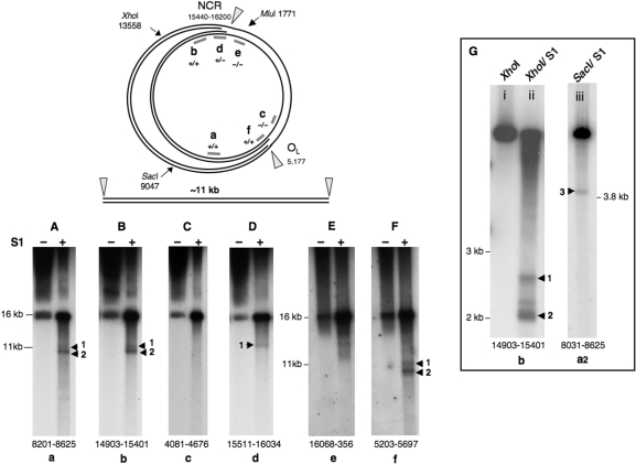
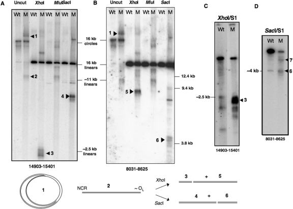
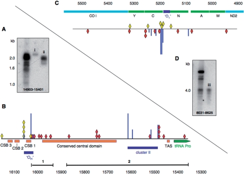
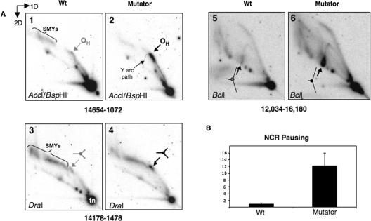
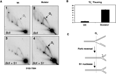
Expression of a proof-reading deficient form of mitochondrial DNA (mtDNA) polymerase gamma, POLG, causes early death accompanied by features of premature ageing in mouse. However, the mechanism of cellular senescence remains unresolved. In addition to high levels of point mutations of mtDNA, the POLG mutator mouse harbours linear mtDNAs. Using one- and two-dimensional agarose gel electrophoresis, we show that the linear mtDNAs derive from replication intermediates and are indicative of replication pausing and chromosomal breakage at the accompanying fragile sites. Replication fork arrest is not random but occurs at specific sites close to two cis-elements known as O(H) and O(L). Pausing at these sites may be enhanced in the case of exonuclease-deficient POLG owing to delayed resumption of DNA replication, or replisome instability. In either case, the mtDNA replication cycle is perturbed and this might explain the progeroid features of the POLG mutator mouse.


Figures

Figure 1.
Figure 1.
Linear 11 kb fragments of mtDNA are released from control mouse liver mtDNA by single-stranded nuclease treatment. The figure shows a schematic diagram of a replication intermediate of mammalian mtDNA, where fork arrest has occurred in the NCR and near OL; nicking by S1, or other, nuclease can in theory release a linear fragment of mtDNA of ∼11 kb as illustrated. S1 nuclease treatment of Balb C mouse liver mtDNA yielded just such fragments based on a series of probes from around the mouse mitochondrial genome (Panels AF). (Panels A–D) represent a single gel: 0.62% agarose, 60 V, 30 h; whereas, the mtDNA of (Panel E) and (Panel F) was separated on a 0.5% agarose gel, at 65 V for 30 h. Treating mouse liver mtDNA with XhoI or SacI, in addition to S1 nuclease, produced shorter fragments (Panel G, i–iii), after separation at 100 V for 4 h on a 0.8% agarose gel. Probes were assigned lowercase letters (a–f), and the numbers at the foot of the gel panels indicate the span of the probes, based on the revised mouse mtDNA reference sequence (17).
Figure 2.
Figure 2.
Linear 11 kb fragments of mtDNA are present at high abundance in mutator mouse liver mtDNA, in the absence of single-strand nuclease treatment. Liver mtDNA samples isolated from wild-type (Wt) and mutator (M) mice were digested with restriction enzymes, XhoI, MluI or SacI, or left uncut, and fractionated by 1D-AGE in TBE buffer. Separation conditions were 55 V for 20 h in 0.4% agarose (Panels A and B) or 100 V for 4 h in 0.8% agarose (Panels C and D). After Southern blotting, membranes were probed with PCR products corresponding to nt 14 903–15 401 (Panels A and C); and nt 8031–8625 (Panels B and D) of mouse mtDNA. The samples in panels (C) and (D) were treated additionally with single-strand specific (S1) nuclease after restriction digestion (see ‘Materials and Methods’ section). The two lanes in panel (C) are different exposures of the same gel; a longer exposure of the digest of Wt mtDNA was needed to show that S1 nuclease generated some fragments of similar size to the abundant short fragments seen in mutator mouse mtDNA digests. The species that distinguished mutator mouse mtDNA from wild-type mtDNA are interpreted as follows: 1—replicating theta structures, or eyebrows, as illustrated at the base of the figure. Wild-type mtDNA samples also include theta structures but their low abundance means they are difficult to detect unless 2D-AGE is applied (12); 2—linear mtDNA fragments of approximately 11 kb; 3XhoI digested mtDNA fragments with one end corresponding to the restriction site at nt 13 558 and the other mapping to the major non-coding region (NCR); 4SacI fragments ∼7 kb, spanning nt 9047 to the NCR; 5—fragments ∼8 kb, with one end close to OL the other nt 13 558; 6—fragments ∼4 kb, with termini at nt 9047 and near OL. The fragments labelled 7 in panel D were gel-extracted, converted to circles, cloned and sequenced, without recourse to S1 nuclease treatment, and found to contain point mutations, which indicated that the novel fragments were the result of SacI site gains (see Supplementary Figure 3 for details).
Figure 3.
Figure 3.
The ends of the prominent linear molecules of mutator mouse mtDNA map to the NCR and in the vicinity of OL. S1 products of mutator mouse mtDNA were recovered from specific regions of agarose gels using Recochips™; repeated separation of some of the material by 1D-AGE (0.8% agarose, 4 h, 100 V) was used to confirm successful recovery (panel A, i and ii; panel D, iii). Fragments were circularized, and the junction amplified, cloned and sequenced. Panel (B) A partial linear map of the mouse mitochondrial genome encompassing the NCR. The ends (red diamonds) identified by direct sequencing were nt 16 033, 16 016, 15 997, 15 995, 15 978, 15 956 (Recochip 1); and 15 875, 15 751, 15 751, 15 750, 15 747, 15 529, 15 489, 15 487, 15 486, 15 467 and 15 389 (Recochip 2). Scarcer ends mapping to the NCR were also cloned and sequenced from control littermates (yellow diamonds). The NCR of mouse mtDNA includes three so-called conserved sequenced boxes (CSBs 1–3), a conserved central domain, and a predicted clover-leaf structure, TAS, which is believed to effect termination of 7S DNA (D-loop) synthesis. Three of the ends (15 751, 15 751 and 15 750) in the conserved central domain are flanked by a GC-rich sequence CCGGGCCC and GGGGG, which is extremely rare in the L-strand of mammalian mtDNAs, suggesting that this may represent a cis-element. Free 5′ ends of DNA identified previously by LM-PCR are represented as vertical blue lines; in panel B the free 5′ ends comprise two clusters, cluster I (OH) and cluster II (12,13,38). The height of the most prominent free end (nt 16 034) was set arbitrarily and the others expressed as a fraction of its height. Panel (C) The sequenced SacI/S1 products with an end mapping close to OL (nt 5160–5191) were nt 4946, 5082, 5133, 5201, 5217, 5255, 5257, 5318, 5477 (red diamonds) in mutator mouse samples and nt 5169, 5193, 5196, 5199, 5244 and 5362 (yellow diamonds) in controls. In humans, free 5′ ends are concentrated in the tRNACys gene (C), which is adjacent to OL (38,39) and free 5′ ends map to similar positions in mouse mtDNA (see Supplementary Figure 2); in panel (C) they are again represented as blue vertical lines. Panel (D) SacI/S1-treated mutator mouse mtDNA analysed by 1D-AGE, iii is the material that was used to define the ends of DNA represented as red diamonds in panel (C).
Figure 4.
Figure 4.
Enhanced replication pausing in the NCR of POLG mutator mouse mtDNA. (A) 2D-AGE analysis of mtDNA of control (WT) and mutator mouse liver was performed after digestion with AccI and BspHI (panels 1 and 2), DraI (panels 3 and 4) or BclI (panels 5 and 6). Arrows indicate the position on a standard Y arc where replication forks frequently arrest [for details of replication fork arrest, see (40)]. Restriction site blockage accounts for the high molecular mass fragments of mtDNA (slow-moving Y-like arcs or SMYs), which were previously attributed to RNA incorporation during mtDNA replication (12). (B) The abundance of paused replication forks mapping to the NCR was 12 times higher than controls based on Typhoon™ phosphorimager (GE Healthcare) quantification of paused RIs. n = 3 experiments using mtDNA derived from two distinct groups of control and mutator mouse livers.
Figure 5.
Figure 5.
Increased replication pausing in the vicinity of OL in mutator mouse mtDNA. (A) Liver mtDNA samples from mutator mouse and controls (WT) were digested with BclI and probed with a PCR product spanning nt 5568–6044 after N2D-AGE, to reveal a fragment encompassing the OL region. (B) Pausing at OL was increased 11-fold based on phosphorimager analysis of gels such as the ones shown in panels 1 and 2. Although increased pausing on the Y arc was more apparent after a brief single-strand (S1) nuclease treatment (1 U for 1 min at 37°C) (panels 3 and 4), quantification indicated that the S1 treatment decreased the relative abundance of paused RIs between POLG mutator mouse and control samples (data not shown), and so the data in B relate to samples that were not treated with S1 nuclease. At least some of the increase in signal on the Y arc produced by S1 nuclease (panel A-4) can be attributed to the removal of single-stranded DNA from regressed forks, as illustrated in (C).

Similar articles

Cited by

References

    1. Li Y, Huang TT, Carlson EJ, Melov S, Ursell PC, Olson JL, Noble LJ, Yoshimura MP, Berger C, Chan PH, et al. Dilated cardiomyopathy and neonatal lethality in mutant mice lacking manganese superoxide dismutase. Nat. Genet. 1995;11:376–381. - PubMed
    1. Reaume AG, Elliott JL, Hoffman EK, Kowall NW, Ferrante RJ, Siwek DF, Wilcox HM, Flood DG, Beal MF, Brown RH, Jr., et al. Motor neurons in Cu/Zn superoxide dismutase-deficient mice develop normally but exhibit enhanced cell death after axonal injury. Nat. Genet. 1996;13:43–47. - PubMed
    1. Schriner SE, Linford NJ, Martin GM, Treuting P, Ogburn CE, Emond M, Coskun PE, Ladiges W, Wolf N, Van Remmen H, et al. Extension of murine life span by overexpression of catalase targeted to mitochondria. Science. 2005;308:1909–1911. - PubMed
    1. Jacobs HT. The mitochondrial theory of aging: dead or alive? Aging Cell. 2003;2:11–17. - PubMed
    1. Bender A, Krishnan KJ, Morris CM, Taylor GA, Reeve AK, Perry RH, Jaros E, Hersheson JS, Betts J, Klopstock T, et al. High levels of mitochondrial DNA deletions in substantia nigra neurons in aging and Parkinson disease. Nat. Genet. 2006;38:515–517. - PubMed

Publication types

Substances