. 2009 May;83(10):5156-67.
doi: 10.1128/JVI.02678-08. Epub 2009 Feb 25.

The 30-amino-acid deletion in the Nsp2 of highly pathogenic porcine reproductive and respiratory syndrome virus emerging in China is not related to its virulence

Affiliations

The 30-amino-acid deletion in the Nsp2 of highly pathogenic porcine reproductive and respiratory syndrome virus emerging in China is not related to its virulence

Lei Zhou et al. J Virol. 2009 May.

Abstract

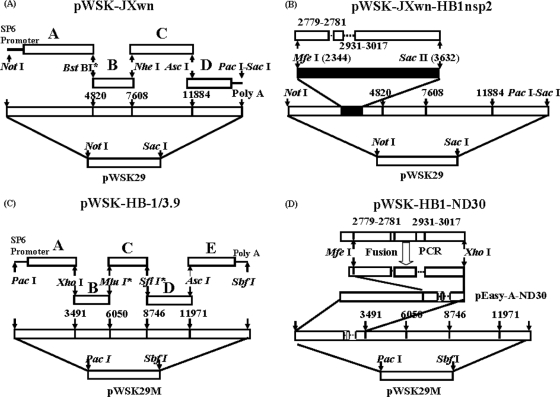
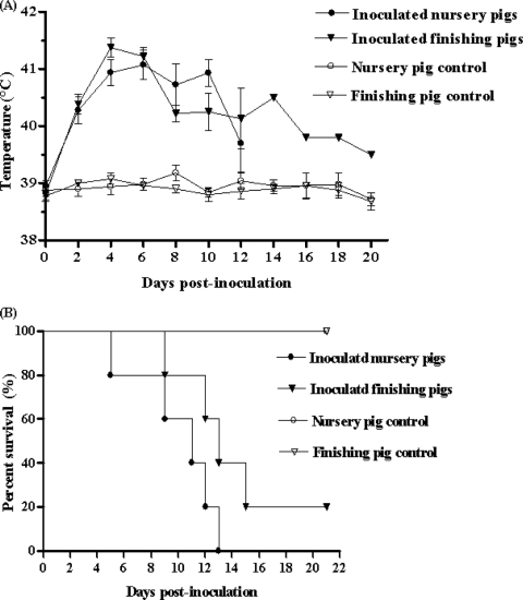
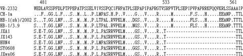
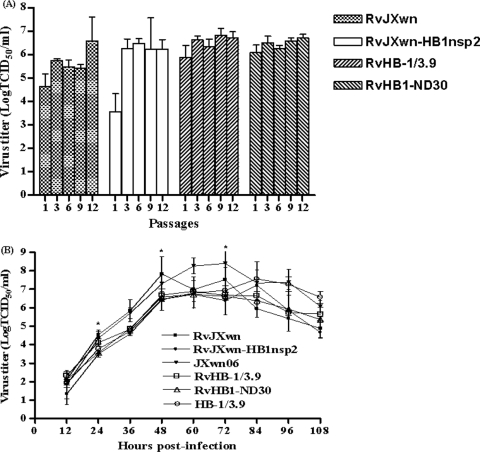
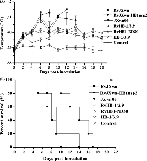
During the past 2 years, an atypical clinical outbreak, caused by a highly pathogenic porcine reproductive and respiratory syndrome virus (PRRSV) with a unique 30-amino-acid deletion in its Nsp2-coding region, was pandemic in China. In this study, we generated four full-length infectious cDNA clones: a clone of the highly virulent PRRSV strain JXwn06 (pWSK-JXwn), a clone of the low-virulence PRRSV strain HB-1/3.9 (pWSK-HB-1/3.9), a chimeric clone in which the Nsp2 region containing the 30-amino-acid deletion was replaced by the corresponding region of the low-virulence PRRSV strain HB-1/3.9 (pWSK-JXwn-HB1nsp2), and a mutated HB-1/3.9 clone with the same deletion in Nsp2 as JXwn06 (pWSK-HB1-ND30). We also investigated the pathogenicities of the rescued viruses (designated RvJXwn, RvJXwn-HB1nsp2, RvHB-1/3.9, and RvHB1-ND30, respectively) in specific-pathogen-free piglets in order to determine the role of the 30-amino-acid deletion in the virulence of the highly pathogenic PRRSV. All the rescued viruses could replicate stably in MARC-145 cells. Our findings indicated that RvJXwn-HB1nsp2 retained high virulence for piglets, like RvJXwn and the parental virus JXwn06, although the survival time of piglets infected with RvJXwn-HB1nsp2 was obviously prolonged. RvHB1-ND30 exhibited low virulence for piglets, like RvHB-1/3.9 and the parental virus HB-1/3.9. Therefore, we conclude that the 30-amino-acid deletion is not related to the virulence of the highly pathogenic PRRSV emerging in China.


Figures

FIG. 1.
FIG. 1.
Strategy for the construction of full-length cDNA clones. (A) Capital letters (A, B, C, D) represent four overlapping fragments amplified from the JXwn06 genome according to the unique restriction enzyme cleavage sites in viral cDNA. A NotI enzyme cleavage site was added to the 5′ end of each fragment, while a 37-nt polyadenosine tail followed by PacI-SacI was added to the 3′ end of the D fragment by PCR mutagenesis. The BstBI site between fragments A and B was created by mutation to be the genetic marker as well as the ligation site. An SP6 promoter with one nontemplated G residue preceded the viral genome. The fragments were inserted into the low-copy-number vector pWSK29 in order from D to A. The completed full-length clone was named pWSK-JXwn. (B) An Nsp2 fragment of 1,378 nt covering the deletion region in JXwn06 was amplified from HB-1/3.9. Two suitable restriction enzyme sites (MfeI and SacII) in fragment A were used to exchange the corresponding fragment of Nsp2. The completed full-length chimeric clone was named pWSK-JXwn-HB1nsp2. (C) Five fragments amplified from HB-1/3.9 were inserted into the modified plasmid pWSK29M to construct the infectious clone. PacI was added to the 5′ end of each fragment, and MluI and SfiI were induced by PCR mutation to ligate the B and C fragments and the C and D fragments. This clone was named pWSK-HB-1/3.9. (D) A discontinuous 90-bp sequence in Nsp2 was deleted by integrating three overlapping segments using fusion PCR, and the product was then cloned into pEasy-A-ND30 using MfeI and XhoI. The modified fragment A, with a 90-bp deletion in the Nsp2 coding region, was finally assembled into the backbone of pWSK-HB-1/3.9 to construct clone pWSK-HB1-ND30.
FIG. 2.
FIG. 2.
Analyses of the pathogenicity of JXwn06. Shown are rectal temperature measurements (A) and survival rates (B) for each group of pigs (n = 5) inoculated with JXwn06. Body temperatures shown are means ± standard deviations (error bars), except for the data from day 14 on for the one inoculated finishing pig that survived.
FIG. 3.
FIG. 3.
Alignment of amino acids in the Nsp2 deletion region of JXwn06 with the sequences of VR-2332 (GenBank accession no. U87392); the earlier Chinese isolates CH-1a (GenBank accession no. AY032626), HB-1(sh)/2002 (GenBank accession no. AY150312), and HB-1/3.9 (GenBank accession no. EU360130); and the highly pathogenic isolates JXA1 (GenBank accession no. EF112445), JX143 (GenBank accession no. EU708726), HuN4 (GenBank accession no. EF635006), and SY0608 (GenBank accession no. EU144079). Dots indicate conserved residues; dashes indicate deleted amino acids at positions 481 and 533 to 561. The deleted amino acid positions were determined based on the genome of VR-2332.
FIG. 4.
FIG. 4.
Identification and differentiation of the rescued viruses. (A) MARC-145 cells infected with third-passage cultures of the viruses were fixed at 48 h postinoculation and examined by immunofluorescence assays using monoclonal antibodies against the N protein (SDOW17) and the Nsp2 protein (E3G11) of PRRSV. McAb, monoclonal antibody. (B) Genetic markers of the rescued viruses were detected by RT-PCR. (C) Partial Nsp2 fragments of the rescued viruses were examined by RT-PCR.
FIG. 5.
FIG. 5.
Stability and growth kinetics of the rescued viruses. (A) The virus titers of the rescued viruses at passages 1, 3, 6, 9, and 12 were determined by microtitration infectivity assays. (B) The growth curves of the four rescued viruses were drawn by assaying the viral titers of the supernatants obtained from 12 h to108 h postinfection by using microtitration infectivity assays. Data are means ± standard deviations (error bars) from three independent trials. Asterisks indicate a significant difference in the viral titer between RvJXwn-HB1nsp2 and RvJXwn or between RvJXwn-HB1nsp2 and JXwn06 (P < 0.05).
FIG. 6.
FIG. 6.
Analyses of the pathogenicities of the rescued viruses for pigs. (A) The rectal temperatures of the inoculated pigs were measured. Data are means ± standard deviations (error bars), except for the temperatures of the one RvJXwn-inoculated pig that survived at 10 and 12 dpi. (B) The survival of the inoculated pigs was recorded.
FIG. 7.
FIG. 7.
Viral loads and antibody kinetics in the sera of pigs inoculated with the rescued viruses. (A) Virus titers were determined by a microtitration infectivity assay. Data are means ± standard deviations (error bars), except for the data representing the one pig inoculated with RvJXwn that survived at 14 dpi. Asterisks indicate significant differences in viral load between RvJXwn-HB1nsp2 and RvJXwn and between RvJXwn-HB1nsp2 and JXwn06 (P < 0.05). (B) An antibody specific for PRRSV N protein was detected using an Idexx Herdchek PRRS 2XR ELISA kit, and the level of antibody was expressed as a sample value/positive value (S/P) ratio. A ratio of ≥0.4 was regarded as indicating seroconversion.

References

    1. Albina, E. 1997. Epidemiology of porcine reproductive and respiratory syndrome (PRRS): an overview. Vet. Microbiol. 55309-316. - PubMed
    1. Allende, R., T. L. Lewis, Z. Lu, D. L. Rock, G. F. Kutish, A. Ali, A. R. Doster, and F. A. Osorio. 1999. North American and European porcine reproductive and respiratory syndrome viruses differ in non-structural protein coding regions. J. Gen. Virol. 80307-315. - PubMed
    1. Bautista, E. M., J. J. Meulenberg, C. S. Choi, and T. W. Molitor. 1996. Structural polypeptides of the American (VR-2332) strain of porcine reproductive and respiratory syndrome virus. Arch. Virol. 1411357-1365. - PubMed
    1. Benfield, D. A., E. Nelson, J. E. Collins, L. Harris, S. M. Goyal, D. Robison, W. T. Christianson, R. B. Morrison, D. E. Gorcyca, and D. W. Chladek. 1992. Characterization of swine infertility and respiratory syndrome (SIRS) virus (isolate ATCC VR-2332). J. Vet. Diagn. Investig. 4127-133. - PubMed
    1. Bøtner, A., J. Nielsen, and V. Bille-Hansen. 1994. Isolation of porcine reproductive and respiratory syndrome (PRRS) virus in a Danish swine herd and experimental infection of pregnant gilts with the virus. Vet. Microbiol. 40351-360. - PubMed

Publication types

MeSH terms

LinkOut - more resources