. 2009 May;83(9):4520-7.
doi: 10.1128/JVI.02601-08. Epub 2009 Feb 25.

Entry of herpes simplex virus 1 and other alphaherpesviruses via the paired immunoglobulin-like type 2 receptor alpha

Affiliations

Entry of herpes simplex virus 1 and other alphaherpesviruses via the paired immunoglobulin-like type 2 receptor alpha

Jun Arii et al. J Virol. 2009 May.

Abstract

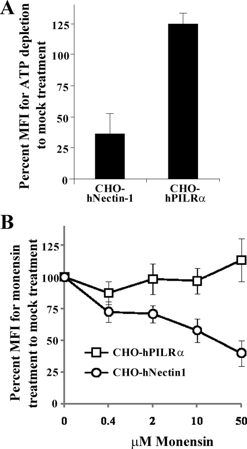
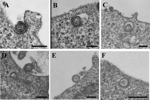
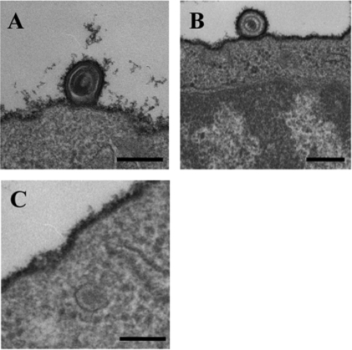
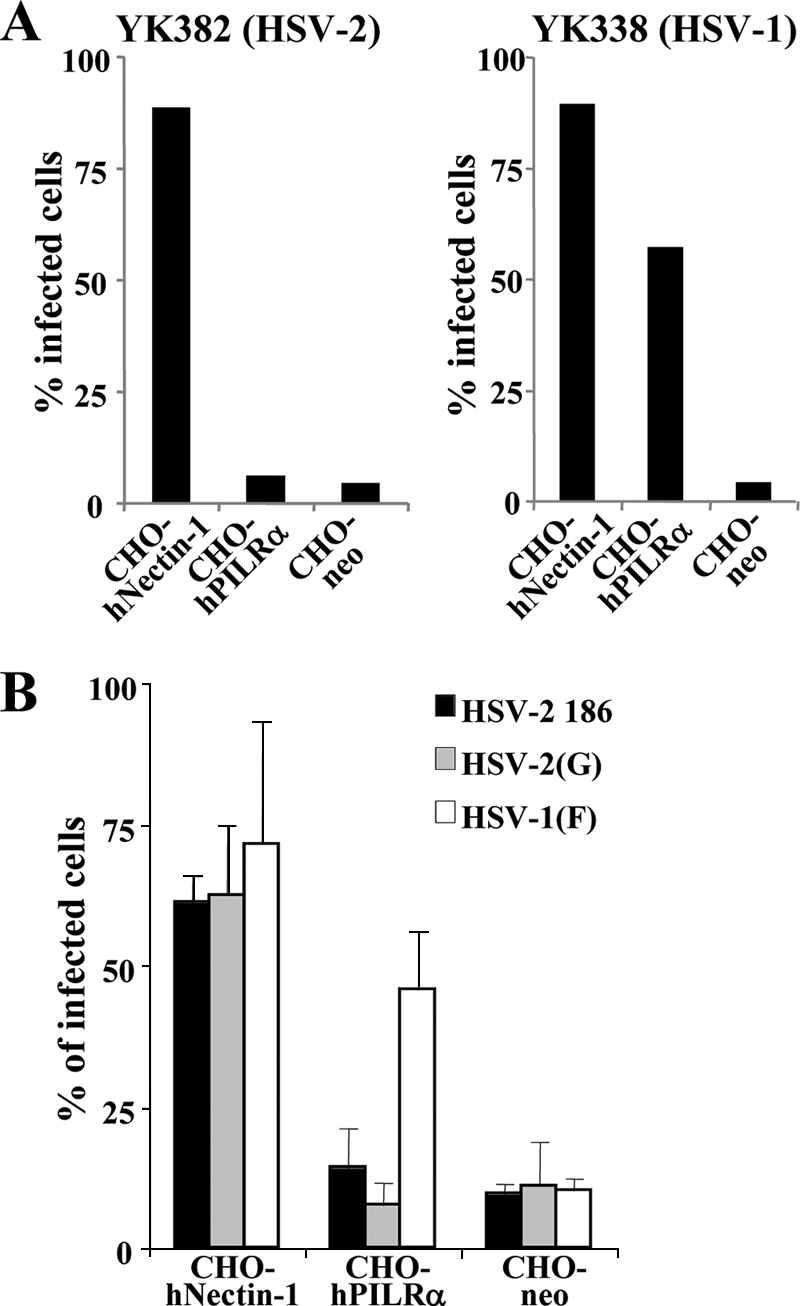
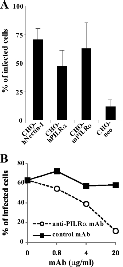
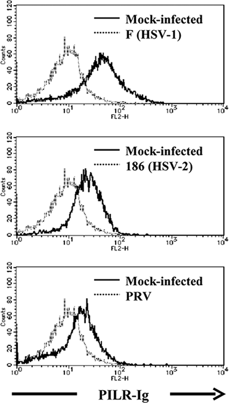
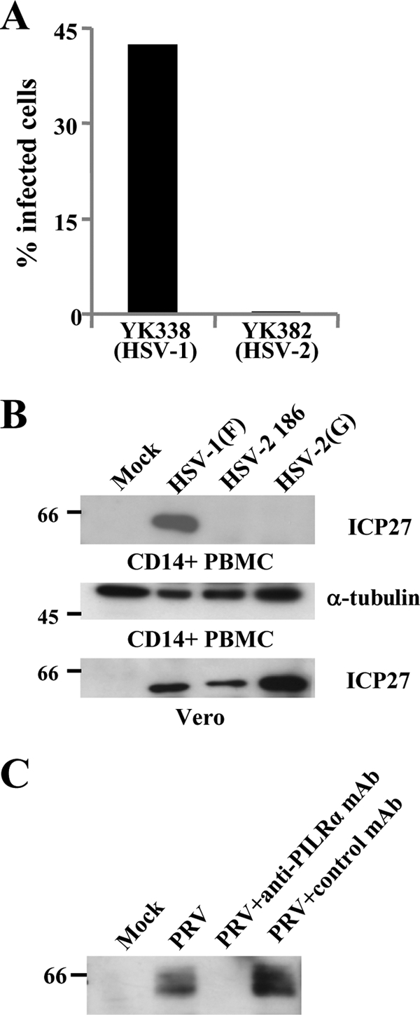
Herpes simplex virus 1 (HSV-1) enters cells either via fusion of the virion envelope and host cell plasma membrane or via endocytosis, depending on the cell type. In the study reported here, we investigated a viral entry pathway dependent on the paired immunoglobulin-like type 2 receptor alpha (PILRalpha), a recently identified entry coreceptor for HSV-1 that associates with viral envelope glycoprotein B (gB). Experiments using inhibitors of endocytic pathways and ultrastructural analyses of Chinese hamster ovary (CHO) cells transduced with PILRalpha showed that HSV-1 entry into these cells was via virus-cell fusion at the cell surface. Together with earlier observations that HSV-1 uptake into normal CHO cells and those transduced with a receptor for HSV-1 envelope gD is mediated by endocytosis, these results indicated that expression of PILRalpha produced an alternative HSV-1 entry pathway in CHO cells. We also showed that human and murine PILRalpha were able to mediate entry of pseudorabies virus, a porcine alphaherpesvirus, but not of HSV-2. These results indicated that viral entry via PILRalpha appears to be conserved but that there is a PILRalpha preference among alphaherpesviruses.


Figures

FIG. 1.
FIG. 1.
Effects of endocytosis inhibitors on HSV-1 entry into cells. (A) CHO-hPILRα and CHO-hNectin-1 cells were infected with YK333 at an MOI of 1 at 4°C for 1 h. The inoculum was then removed, and cells were treated with glucose-free medium containing 2% bovine serum albumin, 0.3% 2-deoxy-d-glucose, and 0.05% sodium azide and held at 4°C for 15 min and then at 37°C for 30 min. Virus that had not penetrated the cells was inactivated by acid treatment, and infection continued for an additional 7 h, after which infected cells were analyzed by flow cytometry. The relative mean fluorescence intensity (MFI) of infected cells treated with energy depletion medium was calculated as the percentage relative to that of mock-treated infected cells. The mean and standard deviation from three independent experiments is shown for each cell type. (B) CHO-hPILRα or CHO-hNectin-1 cells were pretreated with the indicated concentrations of monensin for 1 h at 37°C. Cells were then infected with YK333 at an MOI of 1 for 7 h in the presence of the drug and analyzed by flow cytometry. The relative MFI of infected cells was calculated as the percentage relative to that in the absence of the drug. The mean and standard deviation from three independent experiments is shown for each data point for each cell type.
FIG. 2.
FIG. 2.
EM of HSV-1 entry into CHO cells expressing PILRα. HSV-1(F) was added to CHO-hNectin-1 (A to C) or CHO-hPILRα (D to F) cells for 2 h at 4°C, followed by a shift to 37°C for 2 min and then preparation of the cells for EM. Bar, 200 nm.
FIG. 3.
FIG. 3.
EM of HSV-1 entry into CD14-positive PBMCs. HSV-1(F) was added to primary human CD14-positive PBMCs for 30 min at 4°C, followed by a shift to 37°C for 2 min and then preparation of the cells for EM. Bar, 200 nm.
FIG. 4.
FIG. 4.
HSV-2 infection of CHO cells expressing PILRα. (A) CHO-hNnectin-1, CHO-hPILRα, and CHO-neo cells were infected with HSV-2 YK382 or HSV-1 YK338 at an MOI of 5. At 8 h postinfection, infected cells were divided into two aliquots. One aliquot was examined by fluorescence microscopy (data not shown), and the other was analyzed by flow cytometry (A) to determine the percentages of infected cells. (B) CHO-hNnectin-1, CHO-hPILRα, and CHO-neo cells were infected with wild-type HSV-2 186, HSV-2(G), or HSV-1(F) at an MOI of 5. At 8 h postinfection, infected cells were stained with anti-gD antibody and analyzed by flow cytometry to determine the percentages of infected cells. The mean and standard deviation from three independent experiments is shown for each cell type.
FIG. 5.
FIG. 5.
PRV infection of CHO cells expressing PILRα. (A) CHO-hNectin-1, CHO-hPILRα, CHO-mPILRα, and CHO-neo cells were infected with PRV at an MOI of 5. At 8 h postinfection, infected cells were fixed, permeabilized, stained with anti-EP0 antibody, and analyzed by flow cytometry to determine the percentages of infected cells. The mean and standard deviation from three independent experiments is shown for each cell type. (B) CHO-hPILRα cells were infected with PRV at an MOI of 5 in the presence of various concentrations of anti-PILRα MAb or control MAb. At 8 h postinfection, infected cells were stained with anti-EP0 antibody and analyzed by flow cytometry to determine the percentages of infected cells.
FIG. 6.
FIG. 6.
Binding of PILRα to the cell surface molecule(s) in HSV-1-, HSV-2-, or PRV-infected cells. HEK293T cells were mock infected or infected with wild-type HSV-1(F), HSV-2 186, or PRV at an MOI of 5. At 12 h after infection, infected cells were stained with PILRα-Ig fusion protein and analyzed by flow cytometry.
FIG. 7.
FIG. 7.
HSV-2 and PRV infections of primary human CD14-positive PBMCs. (A) Primary human CD14-positive PBMCs were infected with HSV-1 YK338 or HSV-2 YK382 at an MOI of 5. At 14 h postinfection, infected cells were analyzed by flow cytometry to determine the percentages of infected cells. (B) Primary human CD14-positive PBMCs were infected with wild-type HSV-1(F), HSV-2 186, or HSV-2(G) at an MOI of 5 for 14 h, harvested, and analyzed by immunoblotting with ICP27 or α-tubulin. Vero cells were analyzed by the same procedure as described above and immunoblotted with ICP27. (C) Primary human CD14-positive PBMCs were infected with PRV at an MOI of 5 in the presence (20 μg/ml) or absence of anti-PILRα MAb or control MAb. At 7 h postinfection, infected cells were analyzed by immunoblotting with anti-EP0 antibody. Values to the left of the blots indicate molecular size markers in kilodaltons.

References

    1. Ahmad, R., J. Ennaciri, P. Cordeiro, S. El Bassam, and J. Menezes. 2007. Herpes simplex virus-1 up-regulates IL-15 gene expression in monocytic cells through the activation of protein tyrosine kinase and PKC zeta/lambda signaling pathways. J. Mol. Biol. 36725-35. - PubMed
    1. Atanasiu, D., J. C. Whitbeck, T. M. Cairns, B. Reilly, G. H. Cohen, and R. J. Eisenberg. 2007. Bimolecular complementation reveals that glycoproteins gB and gH/gL of herpes simplex virus interact with each other during cell fusion. Proc. Natl. Acad. Sci. USA 10418718-18723. - PMC - PubMed
    1. Avitabile, E., C. Forghieri, and G. Campadelli-Fiume. 2007. Complexes between herpes simplex virus glycoproteins gD, gB, and gH detected in cells by complementation of split enhanced green fluorescent protein. J. Virol. 8111532-11537. - PMC - PubMed
    1. Bender, F. C., J. C. Whitbeck, H. Lou, G. H. Cohen, and R. J. Eisenberg. 2005. Herpes simplex virus glycoprotein B binds to cell surfaces independently of heparan sulfate and blocks virus entry. J. Virol. 7911588-11597. - PMC - PubMed
    1. Bosnjak, L., M. Miranda-Saksena, D. M. Koelle, R. A. Boadle, C. A. Jones, and A. L. Cunningham. 2005. Herpes simplex virus infection of human dendritic cells induces apoptosis and allows cross-presentation via uninfected dendritic cells. J. Immunol. 1742220-2227. - PubMed

Publication types

MeSH terms

LinkOut - more resources