

Neuregulin signaling is dispensable for NMDA- and GABA(A)-receptor expression in the cerebellum in vivo
- PMID: 19244516
- PMCID: PMC6666233
- DOI: 10.1523/JNEUROSCI.4303-08.2009
Neuregulin signaling is dispensable for NMDA- and GABA(A)-receptor expression in the cerebellum in vivo
Abstract
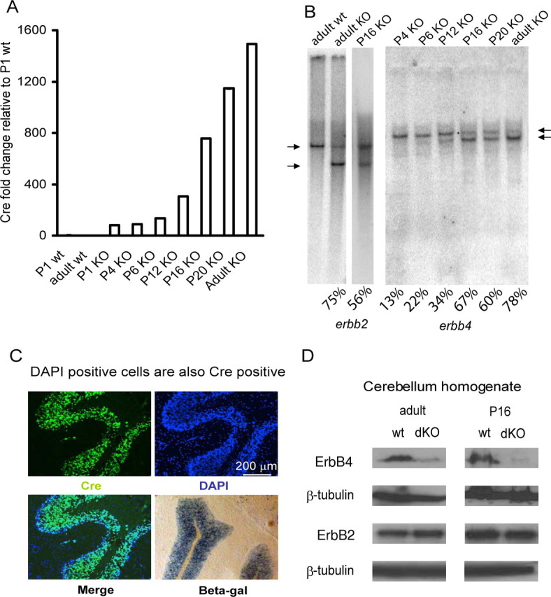
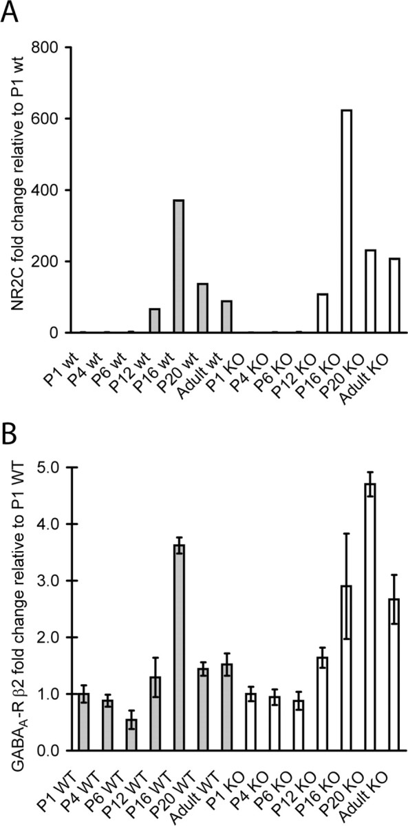
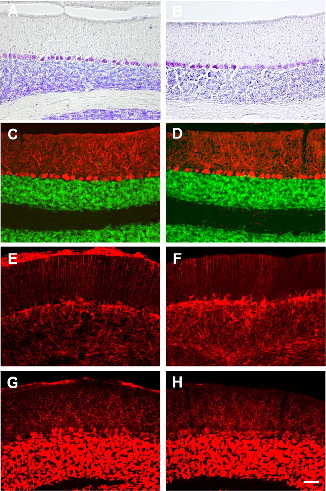
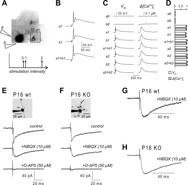
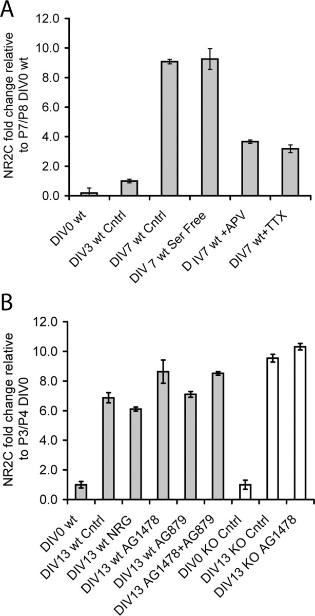
Neuregulin-1s (NRG-1s) are a family of growth and differentiation factors with multiple roles in the development and function in different organs including the nervous system. Among the proposed functions of NRG-1s in the nervous system is the regulation of genes encoding certain neurotransmitter receptors during synapse formation as well as of other aspects of synaptic function. Here, we have examined, in granule cells of the cerebellum in vivo, the role of NRGs in the induction of NMDA receptor (NMDA-R) and GABA(A) receptor (GABA(A)-R), which are thought to be induced by NRG-1 secreted by the synaptic inputs. To this end, we used the Cre/loxP system to genetically ablate the NRG receptors ErbB2 and ErbB4 selectively in these cells, thus eliminating all NRG-mediated signaling to them. Unlike previous reports using cultured granule cells to address the same question, we found that the developmental expression patterns of the mRNAs encoding the NR2C subunit of the NMDA-R and the beta2-subunit of the GABA(A)-R is normal in mice lacking the NRG receptors ErbB2 and ErbB4. Likewise, no alterations in cerebellar morphology nor in certain aspects of cerebellar wiring were resolved in these mutants. We conclude that NRG/ErbB signaling to the granule cells is dispensable for the normal development of their synaptic inputs.
Figures
Similar articles
-
Neuregulin induces GABA(A) receptor subunit expression and neurite outgrowth in cerebellar granule cells.J Neurosci. 1999 Dec 15;19(24):10757-66. doi: 10.1523/JNEUROSCI.19-24-10757.1999. J Neurosci. 1999. PMID: 10594059 Free PMC article.
-
Neuregulin-beta induces expression of an NMDA-receptor subunit.Nature. 1997 Dec 18-25;390(6661):691-4. doi: 10.1038/37795. Nature. 1997. PMID: 9414162
-
NMDA receptor 2 (NR2) C-terminal control of NR open probability regulates synaptic transmission and plasticity at a cerebellar synapse.J Neurosci. 2002 Nov 15;22(22):9687-97. doi: 10.1523/JNEUROSCI.22-22-09687.2002. J Neurosci. 2002. PMID: 12427824 Free PMC article.
-
Neuregulin signaling via erbB receptor assemblies in the nervous system.Mol Neurobiol. 2002 Feb;25(1):67-77. doi: 10.1385/MN:25:1:067. Mol Neurobiol. 2002. PMID: 11890458 Review.
-
The neuregulin-I/ErbB signaling system in development and disease.Adv Anat Embryol Cell Biol. 2007;190:1-65. Adv Anat Embryol Cell Biol. 2007. PMID: 17432114 Review.
Cited by
-
ERBB3-mediated regulation of Bergmann glia proliferation in cerebellar lamination.Development. 2015 Feb 1;142(3):522-32. doi: 10.1242/dev.115931. Epub 2015 Jan 6. Development. 2015. PMID: 25564653 Free PMC article.
-
Neuregulin-1β prevents Ca(2+) overloading and apoptosis through PI3K/Akt activation in cultured dorsal root ganglion neurons with excitotoxicity induced by glutamate.Cell Mol Neurobiol. 2011 Nov;31(8):1195-201. doi: 10.1007/s10571-011-9721-2. Epub 2011 Jun 14. Cell Mol Neurobiol. 2011. PMID: 21671003 Free PMC article.
-
Building better brains: the pleiotropic function of neurotrophic factors in postnatal cerebellar development.Front Mol Neurosci. 2023 May 12;16:1181397. doi: 10.3389/fnmol.2023.1181397. eCollection 2023. Front Mol Neurosci. 2023. PMID: 37251644 Free PMC article. Review.
-
Neuregulin 1 regulates pyramidal neuron activity via ErbB4 in parvalbumin-positive interneurons.Proc Natl Acad Sci U S A. 2010 Jan 19;107(3):1211-6. doi: 10.1073/pnas.0910302107. Epub 2009 Dec 29. Proc Natl Acad Sci U S A. 2010. PMID: 20080551 Free PMC article.
-
Studying Cerebellar Circuits by Remote Control of Selected Neuronal Types with GABA(A) Receptors.Front Mol Neurosci. 2009 Dec 11;2:29. doi: 10.3389/neuro.02.029.2009. eCollection 2009. Front Mol Neurosci. 2009. PMID: 20076763 Free PMC article.
References
-
- Adcock KH, Brown DJ, Shearer MC, Shewan D, Schachner M, Smith GM, Geller HM, Fawcett JW. Axon behaviour at Schwann cell - astrocyte boundaries: manipulation of axon signalling pathways and the neural adhesion molecule L1 can enable axons to cross. Eur J Neurosci. 2004;20:1425–1435. - PubMed
-
- Agresti A, Coull BA. Approximate is better than “Exact” for interval estimation of binomial proportions. Am Stat. 1998;52:119–126.
-
- Aller MI, Jones A, Merlo D, Paterlini M, Meyer AH, Amtmann U, Brickley S, Jolin HE, McKenzie AN, Monyer H, Farrant M, Wisden W. Cerebellar granule cell Cre recombinase expression. Genesis. 2003;36:97–103. - PubMed
-
- Altman J. Postnatal development of the cerebellar cortex in the rat. III. Maturation of the components of the granular layer. J Comp Neurol. 1972;145:465–513. - PubMed
Publication types
MeSH terms
Substances
LinkOut - more resources
Full Text Sources
Molecular Biology Databases
Research Materials
Miscellaneous