. 2009 Feb 25;29(8):2453-66.
doi: 10.1523/JNEUROSCI.4524-08.2009.

The role of Sonic hedgehog of neural origin in thalamic differentiation in the mouse

Affiliations

The role of Sonic hedgehog of neural origin in thalamic differentiation in the mouse

Nora-Emöke Szabó et al. J Neurosci. .

Abstract

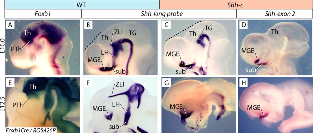
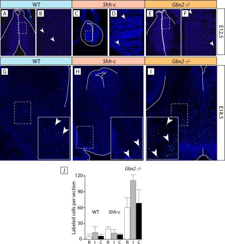
The specification of the intricate neuronal assemblies that characterize the forebrain is not well understood. The ventral spinal cord is specified through a concentration gradient of Sonic hedgehog (Shh) protein secreted by the notochord. Shh is expressed also in the forebrain neuroepithelium (neural Shh) and the underlying notochord and prechordal plate. Neural Shh is essential for the development of the prethalamus (ventral thalamus), but its effects on the thalamus (dorsal thalamus) are still unclear. We hypothesized that neural Shh would act on a previously regionalized dorsal diencephalic region to promote the emergence of specific thalamic nuclear and histological traits. To find out, we generated a conditional mouse mutant line specifically lacking Shh expression in the diencephalic neuroepithelium. We show that the transcription factor Gbx2, required for thalamic development downstream Shh, is expressed in our mutant in a restricted thalamic region and is necessary and sufficient for the differentiation of the medial and intralaminar thalamic nuclei. In the rest of the thalamus, neural Shh is required to promote neuronal aggregation into nuclei as well as axonal extension. In this way, the individual thalamic nuclei show differential dependence on Shh, Gbx2, or both for their differentiation. Additionally, Gbx2 is required for the survival of thalamic neurons.


Figures

Figure 1.
Figure 1.
Abolition of functional Shh expression in the Shh-c caudal diencephalon. A–D, F–H, Whole-mount in situ hybridization for the genes and genotypes indicated. E, Foxb1 lineage mapping by β-galactosidase detection in Foxb1Cre/ROSA26R heterozygotes. A, B, At E10.0, Foxb1 expression (A) overlaps with the incipient ZLI as labeled by Shh (B). C, D, In the Shh-c mutant, Shh transcription is not active in the ZLI but in the diencephalic tegmentum (C). An exon 2 probe shows that all diencephalic Shh mRNA at this age is nonfunctional (D). The dotted lines in B and C show that the mutant thalamus is smaller. E, F, At E12.5, Foxb1 lineage labeling is present in most of the thalamic region (E), overlapping thalamus and ZLI as labeled by Shh (F). The Shh-c mutant shows almost no Shh transcriptional activation in the caudal diencephalon (G) and no functional Shh at all (H). For the abbreviations used in the figures, see Table 1.
Figure 2.
Figure 2.
Thalamic regionalization in Shh-c mutants. Whole-mount in situ hybridization of hemisected E12.5 mouse brains, probes, and genotypes as indicated. A, B, Lack of Ptch1 expression indicates that the Shh signaling pathway is abolished in the Shh-c mutant diencephalon. C–F, The ZLI has disappeared in the Shh-c mutant, as determined by expression of Nkx2-2 and Lhx1. G, H, Thalamic expression of Dbx1 is preserved in the mutant. I, J, Gbx2 is expressed in a small restricted thalamic domain at E12.5 in the Shh-c mutant (arrow in J).
Figure 3.
Figure 3.
Marker expression in the E18.5 wild-type thalamus. A–T, In situ hybridization on E18.5 mouse brain sections for five marker genes as indicated on top of each column. Rows show sections at given rostrocaudal levels, as indicated on the left side, labeled for each of the five markers. Columns correspond to four rostrocaudal levels labeled with the same probe. Some structures have been outlined for clarity. For details, see Results.
Figure 4.
Figure 4.
Marker expression in the E18.5 Shh-c thalamus. A–T, In situ hybridization on E18.5 mouse brain sections for five marker genes as indicated on top of each column. Rows show sections at a given rostrocaudal level, as indicated on the left side, labeled for each of the five markers. Columns correspond to four rostrocaudal levels labeled with the same probe. The arrows in J and O mark continuous rostrocaudal expression of Cdh6 in the central pronucleus. Some structures have been outlined for clarity. For details, see Results.
Figure 5.
Figure 5.
No thalamic axons reach the cortex in the Shh-c brain. Detection of alkaline phosphatase activity in transverse sections of wild-type (A), Shh-c (B), and Gbx2−/− (C) brains at E18.5. The wild-type cortex (A) shows abundant labeled thalamic axons in the cortex, whereas the Shh-c (B) and the Gbx2−/− (C) cortices are completely unlabeled.
Figure 6.
Figure 6.
Marker expression in the E18.5 Gbx2 thalamus. A–T, In situ hybridization on E18.5 mouse brain sections for five marker genes as indicated on top of each column. Rows show sections at a given rostrocaudal level, as indicated on the left side, labeled for each of the five markers. Columns correspond to four rostrocaudal levels labeled with the same probe. The arrows in B, C, G, H, L, and M indicate marker expression in morphologically abolished medial pronucleus. Some structures have been outlined for clarity. For details, see Results.
Figure 7.
Figure 7.
Differentiation markers in the thalamus. In situ detection of mRNA on transverse sections of E12.5 (A–O) or E18.5 (P–U) mouse brains for the genes and genotypes indicated. At E12.5, expression of Olig3 (A–C), Neurog1 (D–F), Neurog2 (G–I), Neurod1 (J–L), and Neurod4 (M–O) is maintained in the mutants. At E18.5, expression of pan-neuronal marker gene Tubb3 (P–R) and astroglial marker gene Gfap (S–U) does not show changes in the mutant brains. In P, Q, S, and T, the thalamus has been outlined for clarity.
Figure 8.
Figure 8.
BrdU labeling in the mutant thalamus. A–C, BrdU-labeled sections of E12.75 thalamus of the three genotypes as indicated. White arrows indicate the labeled thalamic neuroepithelium. D, E, Results from counting absolute numbers of labeled cells (D) and labeling index (E) in the E12.75 thalamic neuroepithelium (±SD) after 3 h. survival. No statistically significant differences were detected. F–I, BrdU-labeled cells (injection at E12.5, collection at E18.5) in the wild-type (F, H) and Shh-c (G, I) thalamus. The thalamus has been outlined in white. Rostrally (F, G), the wild type showed labeled cells at every mediolateral level, whereas in the Shh-c (G), they were confined to a band surrounding the ventricle (dotted line), and the mantle layer (arrow) showed sparse labeling. More caudally (H, I), most labeled cells in the Shh-c (I) did not progress beyond intermediate mediolateral levels (dotted line).
Figure 9.
Figure 9.
Apoptosis in the thalamus of the Gbx2 mutant. A–F, TUNEL of apoptotic cells in the E12.5 thalamus of the three genotypes as indicated. B, D, and F show magnified views of the frames in A, C, and E. Arrowheads indicate apoptotic cells. G–I, Sections through intermediate rostrocaudal levels of the E18.5 thalamus of the three genotypes showing apoptosis detection. The frame in dotted line is shown magnified as inset in each picture. Some apoptotic cells are indicated by arrowheads. J, Mean ± SD of apoptotic cells per histological section of the E18.5 thalamus at rostral (R), intermediate (I), and caudal (C) levels in the three genotypes. The increase at every level in the Gbx2 mutant is significant with respect to the other two genotypes (p < 0.005).
Figure 10.
Figure 10.
Summary of results. A, Involvement of Shh, Gbx2 and unknown factors (X, Y) in the differentiation of thalamic nuclear groups inferred from the mutant phenotypes. The Gbx2 label in a closed box symbolizes restricted default expression of Gbx2. We include the intralaminar nuclei in the medial pronucleus (see Results). Color code as in B. B, Color code for this figure. C, Diagram showing the thalamic pronuclei in four rostrocaudal levels labeled according to the severity of their phenotype in the Shh-c and Gbx2 mutants. D, Thalamic nuclear groups mapped on a flat map (Swanson, 1992) and labeled according to dependence on Shh and/or Gbx2. Each of the three groups encompasses contiguous nuclei. E, Localization of the thalamic neuroepithelium (top) and possible arrangements of the three presumptive regions that will give rise to the different nuclear groups (bottom). Dotted line, Anteroposterior axis of thalamic primordium. C, Central; D, dorsal; M, medial; ETh, epithalamus.

Similar articles

Cited by

References

    1. Alcántara S, Frisén J, del Río JA, Soriano E, Barbacid M, Silos-Santiago I. TrkB signaling is required for postnatal survival of CNS neurons and protects hippocampal and motor neurons from axotomy-induced cell death. J Neurosci. 1997;17:3623–3633. - PMC - PubMed
    1. Altman J, Bayer SA. Development of the rat thalamus: I. Mosaic organization of the thalamic neuroepithelium. J Comp Neurol. 1988;275:346–377. - PubMed
    1. Alvarez-Bolado G, Zhou X, Voss AK, Thomas T, Gruss P. Winged helix transcription factor Foxb1 is essential for access of mammillothalamic axons to the thalamus. Development. 2000;127:1029–1038. - PubMed
    1. Alvarez-Bolado G, Eichele G. Analysing the developing brain transcriptome with the GenePaint platform. J Physiol. 2006;575:347–352. - PMC - PubMed
    1. Andersson E, Tryggvason U, Deng Q, Friling S, Alekseenko Z, Robert B, Perlmann T, Ericson J. Identification of intrinsic determinants of midbrain dopamine neurons. Cell. 2006;124:393–405. - PubMed

Publication types

MeSH terms

LinkOut - more resources