. 2009 Feb 25;29(8):2519-27.
doi: 10.1523/JNEUROSCI.3923-08.2009.

Glycine inhibitory dysfunction induces a selectively dynamic, morphine-resistant, and neurokinin 1 receptor- independent mechanical allodynia

Affiliations

Glycine inhibitory dysfunction induces a selectively dynamic, morphine-resistant, and neurokinin 1 receptor- independent mechanical allodynia

Loïs S Miraucourt et al. J Neurosci. .

Abstract

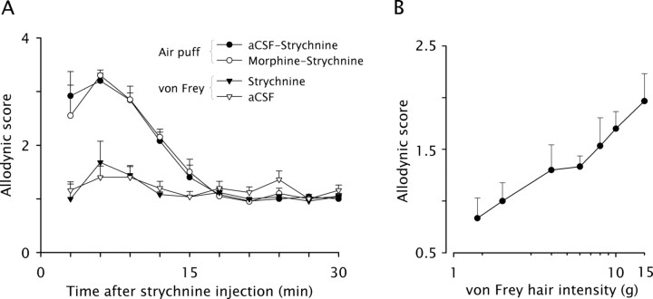
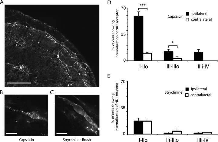
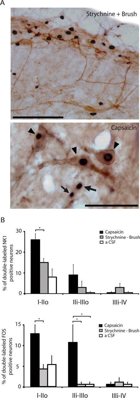
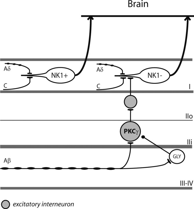
Dynamic mechanical allodynia is a widespread and intractable symptom of neuropathic pain for which there is a lack of effective therapy. We recently provided a novel perspective on the mechanisms of this symptom by showing that a simple switch in trigeminal glycine synaptic inhibition can turn touch into pain by unmasking innocuous input to superficial dorsal horn nociceptive specific neurons through a local excitatory, NMDA-dependent neural circuit involving neurons expressing the gamma isoform of protein kinase C. Here, we further investigated the clinical relevance and processing of glycine disinhibition. First, we showed that glycine disinhibition with strychnine selectively induced dynamic but not static mechanical allodynia. The induced allodynia was resistant to morphine. Second, morphine did not prevent the activation of the neural circuit underlying allodynia as shown by study of Fos expression and extracellular-signal regulated kinase phosphorylation in dorsal horn neurons. Third, in contrast to intradermal capsaicin injections, light, dynamic mechanical stimuli applied under disinhibition did not produce neurokinin 1 (NK1) receptor internalization in dorsal horn neurons. Finally, light, dynamic mechanical stimuli applied under disinhibition induced Fos expression only in neurons that did not express NK1 receptor. To summarize, the selectivity and morphine resistance of the glycine-disinhibition paradigm adequately reflect the clinical characteristics of dynamic mechanical allodynia. The present findings thus reveal the involvement of a selective dorsal horn circuit in dynamic mechanical allodynia, which operates through superficial lamina nociceptive-specific neurons that do not bear NK1 receptor and provide an explanation for the differences in the pharmacological sensitivity of neuropathic pain symptoms.


Figures

Figure 1.
Figure 1.
Glycine disinhibition induces morphine-resistant dynamic, but not static, mechanical allodynia. A, The time course of changes in behavioral responses evoked by dynamic mechanical stimuli (air puff) applied on the face of intracisternally strychnine injected rats is shown. Rats were preemptively injected into the cisterna magna with either morphine or aCSF 30 min before strychnine. The time course of changes in behavioral responses evoked by static mechanical stimuli (6.0 g von Frey hair) applied on the face of intracisternally aCSF or strychnine injected rats is also shown. Graphs represent the mean response (±SEM) of rats (n = 4–5/group) to 5 stimuli applied at 10 s intervals. B, Intensity-response relationship for different von Frey hairs near the aversive threshold. Graph represents the mean response (±SEM) of rats (n = 6) to 5 stimuli applied at 10 s intervals.
Figure 2.
Figure 2.
GABAA receptor-dependent disinhibition induces cardiovascular responses that reflect static, but not dynamic, allodynia. The time course of changes in arterial blood pressure responses (ΔMAP) evoked by dynamic (light brushing) or static (6.0 g von Frey hair) mechanical stimuli applied on the face of anesthetized rats immediately after microinfusion of bicuculline within the MDH is shown. Graphs represent the mean response (±SEM) of rats (n = 4/group) to 10 s long stimuli. Bicuculline significantly and reversibly increased blood pressure in response to face stimulation with a 6 g von Frey for 25 min after application.
Figure 3.
Figure 3.
Morphine does not prevent the activation of the neural circuit underlying dynamic mechanical allodynia. A, Images of Fos-positive cell nuclei and phospho-ERK immunolabeling in the MDH of control (Vehicle) and morphine injected rats. Fos expression and ERK phosphorylation were induced by light brushing of the ipsilateral lip after intracisternal strychnine; subcutaneous morphine (4 mg · kg−1) or vehicle (isotonic saline) were preemptively administered 25 min before strychnine; scale bar, 50 μm. On the left, the approximate location at which images were taken is indicated on the schematic diagram of a coronal section of the MDH. B, Bar histograms summarizing mean Fos expression and ERK phosphorylation in the different laminae of the ipsilateral MDH after light brushing of the lip after intracisternal injection of strychnine or aCSF in vehicle and morphine injected animals (n = 3–4/group). A, B, I–IIo, Laminae I and outer II; IIi–IIIo, laminae inner II and outer III; IIIi–IV, laminae inner III and IV. **p < 0.01.
Figure 4.
Figure 4.
Unlike capsaicin, light, dynamic mechanical stimuli applied under glycine disinhibition do not produce NK1 receptor internalization in dorsal horn neurons. A, Image of NK1 receptor-expressing neurons in the MDH as revealed by immunofluorescence. B, Numerical image illustrating a lamina I MDH neuron internalizing NK1 receptors in response to injection of capsaicin in the upper lip. C, Numerical image illustrating a lamina I NK1 immunopositive neuron in the MDH of a rat in which light brushing of the ipsilateral lip was applied after intracisternal strychnine. Scale bars: A, 200 μm; B, C, 10 μm. D, Bar histograms summarizing the percentage of neurons internalizing the NK1 receptor in the different laminae of the MDH after capsaicin injection into the lip. E, Bar histograms summarizing the percentage of neurons internalizing the NK1 receptor in the different laminae of the MDH after brushing of the lip under strychnine. D, E, I–IIo, Laminae I and outer II; IIi–IIIo, laminae inner II and outer III; IIIi–IV, laminae inner III and IV; *p < 0.05, ***p < 0.001.
Figure 5.
Figure 5.
Rare NK1 receptor-expressing dorsal horn neurons are activated by brushing under glycine disinhibition compared with capsaicin injection. A, Images of Fos-positive cell nuclei (black nuclear staining) and NK1 receptor-expressing neurons (brown staining) in lamina I–III of the MDH. Top, After brushing under glycine disinhibition; scale bar, 100 μm; bottom, after capsaicin injection, where clear examples of double-labeled neurons (arrowheads) and Fos-positive cells that are NK1 receptor negative (arrows) are illustrated; scale bar, 20 μm. B, Bar histograms summarizing the percentages of NK1 receptor-expressing neurons and Fos-positive neurons that are double-labeled in the different laminae of the MDH (n = 3–4/group). Fos expression was induced by light brushing of the ipsilateral lip after intracisternal strychnine or by capsaicin injection into the upper ipsilateral lip. Control animals (aCSF) received intracisternal aCSF and brushing without strychnine. I–IIo, Laminae I and outer II; IIi–IIIo, laminae inner II and outer III; IIIi–IV, laminae inner III and IV; *p < 0.05.
Figure 6.
Figure 6.
Schematic diagram illustrating the current understanding of the dorsal horn neural circuit involved in dynamic mechanical allodynia. PKCγ neurons that receive innocuous input via myelinated afferent fibers are part of a local excitatory circuit that mediates dynamic mechanical allodynia after loss of glycinergic inhibitory control (Miraucourt et al., 2007). The exact location of the glycinergic interneurons (GLY) involved in the inhibitory control of the circuit is not known. Although the target of the PKCγ interneurons is still a matter of investigation (Neumann et al., 2008), the current results provide evidence that the dorsal horn output neurons of the circuit are lamina I neurons that lack the NK1 receptor (NK1).

References

    1. Ahmadi S, Lippross S, Neuhuber WL, Zeilhofer HU. PGE(2) selectively blocks inhibitory glycinergic neurotransmission onto rat superficial dorsal horn neurons. Nat Neurosci. 2002;5:34–40. - PubMed
    1. Allen BJ, Li J, Menning PM, Rogers SD, Ghilardi J, Mantyh PW, Simone DA. Primary afferent fibers that contribute to increased substance P receptor internalization in the spinal cord after injury. J Neurophysiol. 1999;81:1379–1390. - PubMed
    1. Baccei M, Fitzgerald M. Development of pain pathways and mechanisms. In: McMahon SB, Koltzenburg M, editors. Wall and Melzack's textbook of pain. Oxford: Churchill Livingstone; 2006. pp. 143–158.
    1. Baron R. Mechanisms of disease: neuropathic pain–a clinical perspective. Nat Clin Pract Neurol. 2006;2:95–106. - PubMed
    1. Besse D, Lombard MC, Zajac JM, Roques BP, Besson JM. Pre- and postsynaptic distribution of mu, delta and kappa opioid receptors in the superficial layers of the cervical dorsal horn of the rat spinal cord. Brain Res. 1990;521:15–22. - PubMed

Publication types

MeSH terms

LinkOut - more resources