. 2009 Feb 25;29(8):2597-610.
doi: 10.1523/JNEUROSCI.5439-08.2009.

The neuropeptide pigment-dispersing factor adjusts period and phase of Drosophila's clock

Affiliations

The neuropeptide pigment-dispersing factor adjusts period and phase of Drosophila's clock

Taishi Yoshii et al. J Neurosci. .

Abstract

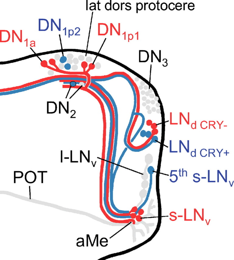
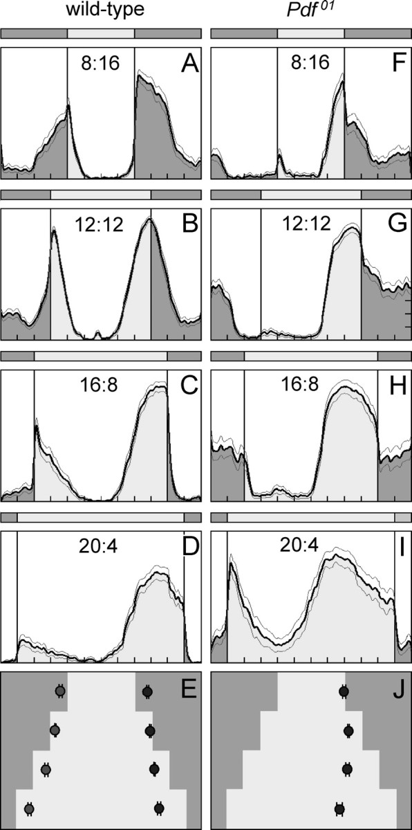
The neuropeptide pigment-dispersing factor (PDF) is a key transmitter in the circadian clock of Drosophila melanogaster. PDF is necessary for robust activity rhythms and is thought to couple the circadian oscillations of the clock neurons. However, little is known about the action of PDF on individual clock neurons. Here, we combined the period-luciferase reporter system with immunolabeling of clock proteins in wild-type and Pdf(01) mutants to dissect the effects of PDF on specific subgroups of clock neurons. Additionally, PDF levels were elevated to higher than normal levels using specific neural mutants, and a correlation analysis of locomotor activity and clock protein staining served to determine the periods of specific clock cells. We found that PDF has multiple effects on the clock neurons: In some groups of clock neurons, PDF was required for maintaining the oscillations of individual cells, and in others, PDF was required for synchronous cycling of the individual members. Other clock neurons cycled with high amplitude in absence of PDF, but PDF affected their intrinsic clock speed. Sometimes PDF shortened and sometimes PDF lengthened period. Our observations indicate that PDF is crucial for adjusting cycling amplitude, period, and phase of the different players in the circadian clock. Under natural conditions PDF may be required for adapting Drosophila's clock to varying photoperiods. Indeed, we show here that Pdf(01) mutants are not able to adapt their activity to long photoperiods in a wild-type manner.


Figures

Figure 1.
Figure 1.
Circadian clock neurons in the right brain hemisphere of the fruit fly. PDF is expressed in two groups of the lateral neurons, the l-LNv and s-LNv. Of these neurons, the arborization patterns are shown; all others are omitted for clarity. The fifth PDF-negative s-LNv is located among the l-LNv, and the LNd are located dorsally of the l-LNv. Whereas all s-LNv and l-LNv cells contain CRY, only three of the six LNd are CRY-positive. The dorsal neurons in the posterior dorsal brain were traditionally divided in three groups (DN1, DN2, and DN3), but they seem to be composed of heterologous cells. Among the DN1, two cells are located in more anterior regions and are therefore called DN1anterior (DN1a), whereas the other up to 15 DN1 are called DN1posterior (DN1p). The DN1a and six of the DN1p cells are CRY-positive. The two DN2 cells are located close to the s-LNv terminals and both are CRY-negative. The DN3 consist of ∼40 neurons of predominantly small size that are all CRY-negative; but there are few larger neurons among them that might have different properties.
Figure 2.
Figure 2.
Locomotor activity and bioluminescence rhythm of flies carrying the 8.0-luc:9 transgene in the wild-type and the Pdf01 background. Control flies (y w;8.0-luc:9) show a free-running rhythm with an average period of 24.1 h (A), whereas y w;8.0-luc:9;Pdf01 mutants exhibit weak rhythms with an average period of 22.8 h (B) (compare Table 1). C, Averaged bioluminescence rhythm of 45 control flies and 28 Pdf01 mutants. The flies were recorded for 6 d in DD after initially being entrained to three cycles of 12:12 h LD. The y w;8.0-luc:9;Pdf01 flies showed rather strong oscillations with a mean period of 22.7 ± 0.1 h, whereas y w;8.0-luc:9 flies showed more dampened oscillations with a mean period of 23.1 ± 0.1 h. The weak dampening present in the y w;8.0-luc:9;Pdf01 flies is entirely attributable to slightly different free-running periods of individual flies as can be seen in E, in which the oscillations of individual y w;8.0-luc:9;Pdf01 flies are shown (n = 10). In contrast, a dampening of the oscillations occurs already on the level of individual wild-type flies (n = 10) (D), indicating that the oscillating DN and LNd cells become out of phase in the presence of PDF.
Figure 3.
Figure 3.
TIM oscillations in the different clock neurons of wild-type flies and Pdf01 mutants for 5 d in constant darkness. The time axis is in circadian time. Wild-type flies and Pdf01 mutants have different periods (Fig. 2); therefore, a circadian day of wild-type flies is 24.2 h long and a circadian day of Pdf01 mutants is 22.8 h long. For both fly strains, circadian time 23 means 1 h before the beginning of the subjective day, and circadian time 11 means 1 h before the beginning of the subjective night. Usually, clock protein levels are high at circadian time 23 and low at circadian time 11. For wild-type flies, this is true for all CRY-positive neurons: the s-LNv, the fifth s-LNv, three LNd, the DN1a, and six DN1p (for significances, see Table 2). However the CRY-negative LNd seem to cycle with different period and are in antiphase with the CRY-positive neurons on the fifth day in DD. The CRY-negative DN1p and DN2 cycled in antiphase to the CRY-positive neurons already earlier, but lost their significant oscillations at the end of the experiment (Table 2). No significant cycling was seen in the CRY-negative DN3 (except for day 1). Note that, in Pdf01 mutants, the CRY-negative and CRY-positive LNd remain in synchrony with each other; the oscillations in the s-LNv and DN1a slowly dampen, and the oscillations in the CRY-positive DN1p disappear quickly after transfer into DD (Table 2 indicates days at which ANOVA revealed significant oscillations); for additional explanations, see text.
Figure 4.
Figure 4.
Spatial expression pattern of the PER–LUC (luciferase) fusion protein in brains of 8.0-luc:9 transgenics. A–B, Monitoring of the luciferase expression with a high-sensitive camera show luminescent spots in the dorsal and dorsolateral brain of control (A) and Pdf01 (B) brains. For each genotype, four brains are shown. Note that, in the Pdf01 background, the LNd are stronger luminescent than the DN. C–K, To reveal transgene expression at higher resolution per01;8.0-luc:9 brains are triple labeled with anti-PER, anti-TIM, and anti-PDF or with anti-CRY, anti-TIM, and anti-PDF at the peak point of staining (1 h before lights-on). Overlays of all labelings are found in E, H, and K. (The orange arrow in E points to the fiber tract originating from the s-LNv and terminating in the distal dorsal protocerebrum close to the DN1 and DN2). PER labeling is only present in neurons that express the transgene (C, F), whereas endogenous TIM is found in all clock neurons (D, G, J. However, nuclear TIM occurs only in the PER-positive cells (arrowheads point to cells that had cytoplasmic TIM and consequently were PER-negative). C, F, PER labeling shows that the 8.0-luc:9 transgene is expressed in the DN1a, most DN1p, the DN2, most DN3, and in four LNd cells. E, H, K, CRY and TIM labeling show that three of the four PER-positive LNd cells are CRY-negative (the arrowheads in D–K point to the CRY-negative cells that have cytoplasmic TIM). L, M, Transgene expression in the Pdf01 background. Note that the DN cells (L) are much weaker labeled as in the wild-type background, but that all six LNd are strongly labeled (M). Additionally, PER is always present in the fifth s-LNv cell (M).
Figure 5.
Figure 5.
Actograms of so1 and somda mutants and TIM staining in the clock neurons of both mutants at two times on days 3–4 in DD (red and blue points). Ten flies were stained at each time for each mutant, respectively. As can be seen in the example actograms, so1 mutants show one rhythm free-running with long period as soon as released to DD (A), whereas somda mutants reveal two rhythmic components in DD, which free-run with short and long periods, respectively, and that were 180° out of phase at the times of staining (C). B, In so1 mutants, the CRY-positive clock neurons were strongly TIM-immunoreactive in the early subjective morning, just before the flies became active (A, red point; B, red columns) and less stained in the subjective evening, when the flies were most active (A, blue point; B, blue columns). The CRY-negative neurons did not show this difference. ANOVA revealed a significant influence of the time of staining on labeling intensity (F (1,165) = 30.68; p < 0.001) and that this influence was dissimilar in the different neuronal groups (F (7,165) = 3.96; p < 0.001). The post hoc test showed that only the CRY-positive neurons showed significantly higher TIM staining in the early subjective morning compared with the evening. No significant differences were revealed for the CRY-negative neurons. D, In somda mutants, ANOVA showed again a significant influence of time on staining intensity (F (1,179) = 4.62; p = 0.03), which was strongly dissimilar in the different neurons (F (8,179) = 34.78; p < 0.001). No significant staining differences were only found in the CRY-negative DN1p remain. The other neurons were either strongly TIM-immunoreactive at the activity maximum of the long-period component (C, blue point; D, blue columns) or at the activity maximum of the short-period component (C, red point; D, red columns). For additional explanations, see text. The green point in C indicates the time at which short- and long-period components crossed each other and were consequently in phase again. At this day, the different clock neurons cycled in synchrony with each other (supplemental Fig. S1, available at www.jneurosci.org as supplemental material). Error bars indicate SEM.
Figure 6.
Figure 6.
TIM labeling showing the internal desynchronization among the dorsal and lateral neurons in somda mutants stained at the two time points marked by red and blue in Figure 5 (red time point, left panel; blue time point, right panel). Both brains were triple stained with anti-TIM (green), anti-CRY (magenta), and anti-PDF precursor (orange). The large pictures in the center of both panels depict the overlay of all three labelings (combination of 10 confocal stacks). Depending on their amount of TIM and CRY, the PDF-positive LNv cells appear in yellow to orange, neurons that have no PDF (e.g., some LNd and DN1) but equal amounts of TIM and CRY appear in white, those that contain only TIM are in green, and those that contain CRY but no or only tiny amounts of TIM are in magenta. To reveal TIM and CRY staining more clearly, single labeling of TIM and CRY of several clock neurons were enlarged and shown separately above and below the central pictures (DN1–2 and LNd on top, LNv on bottom; see insets in the large pictures). At the “red” time point (left panels), TIM was high in one pair of CRY-positive DN1p (the DN1p2) (Fig. 6) and in one cell of the DN2 (note that the DN2 cells are CRY-negative). No TIM labeling was found in the CRY-positive DN1a, and only weak TIM labeling in the CRY-positive DN1p1 and some CRY-negative DN1p. Among the LNd, TIM was high in the three CRY-positive LNd, and no TIM-staining at all was present in the three CRY-negative LNd. Among the LNv, TIM was high in the fifth s-LNv and in the l-LNv that were not evaluated. Almost no TIM was found in the PDF-positive s-LNv. At the “blue” time point (right panels), TIM was high in the DN1a and the DN1p1. Only low amounts of TIM were found in the remaining DN1p and in the DN2. Among the LNd, the CRY-negative LNd were strongly stained by TIM, whereas the CRY-positive LNd contained only little TIM. Of the LNv, again the l-LNv were strongly stained (not evaluated), but now the PDF-positive s-LNv were highly TIM-immunoreactive and little TIM was present in the fifth s-LNv.
Figure 7.
Figure 7.
Diagram summarizing our present view on the differential action of PDF on the clock neurons in somda mutants (only the right brain hemisphere is shown). Neurons that lengthen their period on excessive amounts of PDF in the dorsal brain are shown in blue. Neurons that shorten their period under the same conditions are shown in red. Neurons not assessed in the present study or neurons with unclear response to PDF are shown in gray. Note that the aberrant arborizations of the l-LNv are omitted for better clarity. The s-LNv, fifth s-LNv, l-LNv, DN1a, and the three CRY-positive LNd neurons arborize in the aMe, in which PDF secreted by the l-LNv cells might serve as coupling factor in wild-type flies (Wülbeck et al., 2008, their Discussion). Furthermore, fibers from the s-LNv, the DN1a, DN1p1, and the CRY-positive and -negative LNd cells may contact each other in the lateral dorsal protocerebrum (lat dors protocere), in which PDF seems to be rhythmically released from the s-LNv cells (Park et al., 2000). The fifth s-LNv cell was also found to project toward the dorsolateral protocerebrum, but it is not clear whether it reached this area or whether it terminated before (Helfrich-Förster et al., 2007). The l-LNv cells do not project into the dorsolateral brain in wild-type flies, but do so extensively in somda mutants (Wülbeck et al., 2008). We suppose that extensive amounts of PDF secreted into this brain region shorten the periods of the CRY-positive DN1a and DN1p1 cells. The latter two groups may feedback on the s-LNv forcing them also to free-run with short period (for details, see supplemental Discussion, available at www.jneurosci.org as supplemental material).
Figure 8.
Figure 8.
Average activity profiles of wild-type flies and Pdf01 mutants under different photoperiods. Photoperiods are indicated in the diagrams, whereby 8:16 means 8 h day and 16 h night; 12:12, 12 h day and 12 h night; 16:8, 16 h day and 8 h night; and 20:4, 20 h day and 4 h night. Each activity profile represents the average of 30 flies. Wild-type flies showed prominent morning and evening activity peaks under all photoperiods. The morning peaks advanced with increasing day length, whereas the evening peaks delayed. As a consequence, the midday trough became larger the longer the day length. This can nicely be seen in the phase plot of morning and evening peaks (±SEM) (E). Pdf01 mutants did not show a clear morning peak. Under LD 8:16 (F) and 20:4 (I), the flies seem to respond to lights-on with an increase of activity, and in LD 16:8 (H), a small peak was present ∼4 h after lights-on; furthermore, an activity bout was present in the middle of the night under all photoperiods except 20:4 (I). None of these peaks could be unequivocally regarded as morning peak, and therefore no average phases of the morning peak was calculated (J). The evening peak was well pronounced in Pdf01 mutants. It occurred earlier than in wild-type flies, and it did not delay with increasing day length (J). According to these results, Pdf01 mutants are unable to avoid the midday heat in long summer days.

Similar articles

Cited by

References

    1. Abrahamson EE, Moore RY. Suprachiasmatic nucleus in the mouse; retinal innervation, intrinsic organization and efferent projections. Brain Res. 2001;916:172–191. - PubMed
    1. Aton SJ, Herzog ED. Come together, right… now: synchronization of rhythms in a mammalian clock. Neuron. 2005;48:531–534. - PMC - PubMed
    1. Aton SJ, Colwell CS, Harmar AJ, Waschek J, Herzog ED. Vasoactive intestinal polypeptide mediates circadian rhythmicity and synchrony in mammalian clock neurons. Nat Neurosci. 2005;8:476–483. - PMC - PubMed
    1. Bachleitner W, Kempinger L, Wülbeck C, Rieger D, Helfrich-Förster C. Moonlight shifts the endogenous clock of Drosophila melanogaster . Proc Natl Acad Sci U S A. 2007;104:3538–3543. - PMC - PubMed
    1. Blanchardon E, Grima B, Klarsfeld A, Chélot E, Hardin PE, Préat T, Rouyer F. Defining the role of Drosophila lateral neurons in the control of circadian rhythms in motor activity and eclosion by targeted genetic ablation and PERIOD protein overexpression. Eur J Neurosci. 2001;13:871–888. - PubMed

Publication types

MeSH terms

LinkOut - more resources