. 2009 May 26;48(20):4331-43.
doi: 10.1021/bi900109z.

Substrate-triggered formation and remarkable stability of the C-H bond-cleaving chloroferryl intermediate in the aliphatic halogenase, SyrB2

Affiliations

Substrate-triggered formation and remarkable stability of the C-H bond-cleaving chloroferryl intermediate in the aliphatic halogenase, SyrB2

Megan L Matthews et al. Biochemistry. .

Abstract

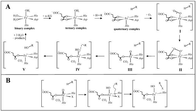
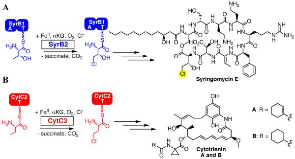
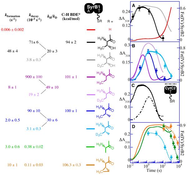
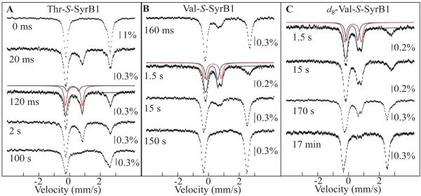
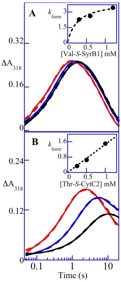
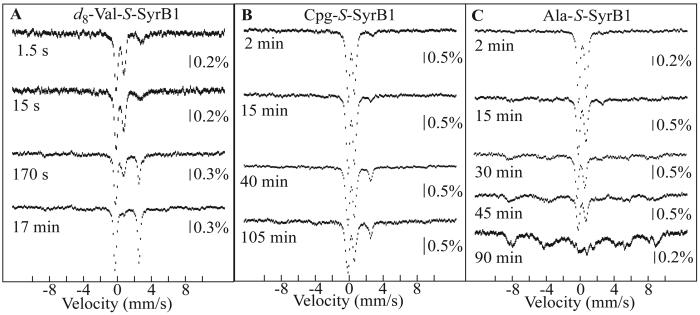
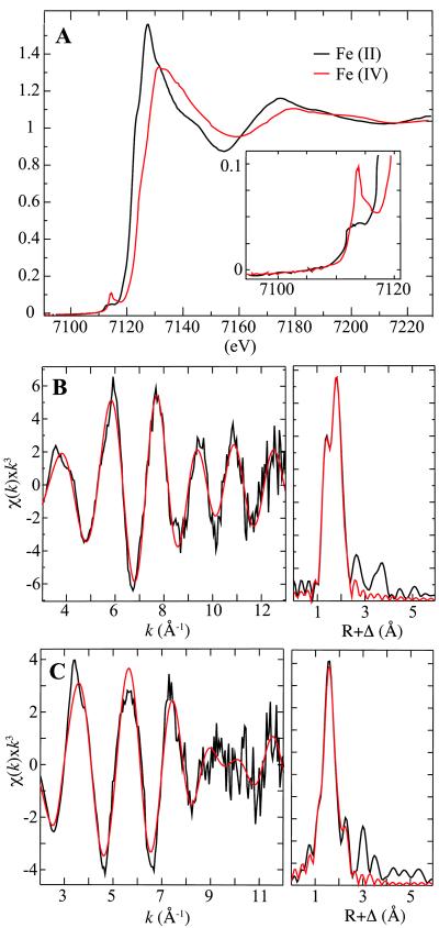
Aliphatic halogenases activate O(2), cleave alpha-ketoglutarate (alphaKG) to CO(2) and succinate, and form haloferryl [X-Fe(IV)O; X = Cl or Br] complexes that cleave aliphatic C-H bonds to install halogens during the biosynthesis of natural products by non-ribosomal peptide synthetases (NRPSs). For the related alphaKG-dependent dioxygenases, it has been shown that reaction of the Fe(II) cofactor with O(2) to form the C-H bond-cleaving ferryl complex is "triggered" by binding of the target substrate. In this study, we have tested for and defined structural determinants of substrate triggering (ST) in the halogenase, SyrB2, from the syringomycin E biosynthetic NRPS of Pseudomonas syringae B301D. As for other halogenases, the substrate of SyrB2 is complex, consisting of l-Thr tethered via a thioester linkage to a covalently bound phosphopantetheine (PPant) cofactor of a carrier protein, SyrB1. Without an appended amino acid, SyrB1 does not trigger formation of the chloroferryl intermediate state in SyrB2, even in the presence of free l-Thr or its analogues, but SyrB1 charged either by l-Thr (l-Thr-S-SyrB1) or by any of several non-native amino acids does trigger the reaction by as much as 8000-fold (for the native substrate). Triggering efficacy is sensitive to the structures of both the amino acid and the carrier protein, being diminished by 5-24-fold when the native l-Thr is replaced with another amino acid and by approximately 40-fold when SyrB1 is replaced with the heterologous carrier protein, CytC2. The directing effect of the carrier protein and consequent tolerance for profound modifications to the target amino acid allow the chloroferryl state to be formed in the presence of substrates that perturb the ratio of its two putative coordination isomers, lack the target C-H bond (l-Ala-S-SyrB1), or contain a C-H bond of enhanced strength (l-cyclopropylglycyl-S-SyrB1). For the latter two cases, the SyrB2 chloroferryl state so formed exhibits unprecedented stability (t(1/2) = 30-110 min at 0 degree C), can be trapped at high concentration and purity by manual freezing without a cryosolvent, and represents an ideal target for structural characterization. As initial steps toward this goal, extended X-ray absorption fine structure (EXAFS) spectroscopy has been used to determine the Fe-O and Fe-Cl distances and density functional theory (DFT) calculations have been used to confirm that the measured distances are consistent with the anticipated structure of the intermediate.


Figures

SCHEME 1
SCHEME 1
HAG mechanism (10) for the Fe(II)- and αKG-dependent hydroxylases (A) and hypothesis for how the hydroxylase and halogenase mechanisms diverge (B) (22).
SCHEME 2
SCHEME 2
Reaction catalyzed by the Fe(II)- and αKG-dependent halogenases SyrB2 (A) and CytC3 (B).
Figure 1
Figure 1
Absorbance-versus-time traces (318 nm) obtained after mixing O2-saturated buffer (20 mM Hepes, pH 7.5) at 5 °C with an equal volume of an O2-free solution containing SyrB2 (360 μM), Fe(II) (300 μM), αKG (10 mM), Cl (100 mM) in absence of substrate (A, red trace) or in presence of the indicated substrate (≥ 750 μM). Substrates include forms of SyrB1 (solid lines) or CytC2 (dashed lines) charged by l-Thr (A and C, black trace), d5-l-Thr (A, gray trace), l-Ala (D, green trace), l-Aba (B, dark pink trace), d6-l-Aba (B, light pink trace), l-Val (B, dark blue trace), d8-l-Val (B, light blue trace) and l-Cpg (D, orange trace). Filled circles indicate the equivalents of total Fe(IV) determined by Mössbauer spectroscopy. For the d8-l-Val, l-Cpg, and l-Ala substrates in D, the data points obtained from the Mössbauer experiments were fit by the equation for an exponential decay (light blue, green, and orange dashed decay traces) to obtain the values of kobs for decay quoted in the text. *The BDEs indicated in the scheme (44) correspond to the target C-H bond strength of small molecules representative of the amino acid side chain: 2-propanol, propane, isobutane, and cyclopropane correspond to the side chains of l-Thr, l-Aba, l-Val, and l-Cpg, respectively.
Figure 2
Figure 2
4.2-K/0-mT Mössbauer spectra of selected samples prepared by reacting the SyrB2•Fe(II)•Cl•αKG•substrate complex at 5 °C with O2-saturated buffer. Substrates and reaction times are indicated. The final sample compositions are given in Materials and Methods. Solid lines are simulations of the spectra of the two Fe(IV) components of the chloroferryl state (red and blue) and the summation of their contributions (black). The isomer shift and quadrupole splitting parameters of the Fe(IV) intermediate(s) are as follows: l-Thr, δ1=0.30 mm/s, ΔEQ,1=1.09 mm/s and δ2=0.23 mm/s, ΔEQ,2=0.76 mm/s; l-Val and d8-l-Val, δ1=0.29 mm/s, ΔEQ,1=1.09 mm/s and δ2=0.24 mm/s, ΔEQ,2=0.73 mm/s.
Figure 3
Figure 3
Dependencies of ST efficacy on the concentrations of two non-native substrates in the reactions of the Fe(II)•SyrB2•αKG•Cl•substrate complex with O2 at 5 °C. A318-versus-time traces from stopped-flow experiments in which the concentration of l-Val-S-SyrB1 (A) or l-Thr-S-CytC2 (B) was varied from 0.30 mM (2-fold excess over the SyrB2•Fe(II)•αKG•Cl complex; black trace), to 0.60 mM (4-fold excess; blue trace) to 1.2 mM (8-fold excess; red trace). The insets show plots of the kobs for formation from regression analysis as a function of [substrate]. In A the data points (filled circles) fit best to a hyperbola characteristic of saturation of SyrB2 by l-Val-S-SyrB1. By contrast, the inset of panel B shows a linear dependence of the kobs for formation on [l-Thr-S-CytC2], indicative of weak binding of the substrate to SyrB2.
Figure 4
Figure 4
4.2-K/53-mT Mössbauer spectra of selected samples collected over ∼ ± 12 mm/s range of Doppler velocities to obtain the decay kinetics of the chloroferryl state formed by reacting the SyrB2•Fe(II)•Cl•αKG•substrate complex at 5 °C with O2. Substrates and reaction times are indicated. The final compositions of the samples are given in Materials and Methods.
Figure 5
Figure 5
Fe K-edge x-ray absorption data for the SyrB2•Fe(II)•αKG•Cll-Cpg-CytC2 sample (A, black trace) and the chloroferryl state resulting from reaction of this complex with O2 (A, red trace). The pre-edge region is expanded in the inset. EXAFS data for the chloroferryl state (B, left panel) and Fourier transform of the EXAFS data (B, right panel). The raw data are shown in black and the best fits in red. The 57Fe Mössbauer spectrum of the same sample is given in the Supporting Information (Figure S1, spectrum G in Supporting Information). EXAFS data (C, left panel) for the Fe(II) reactant complex and the Fourier transform of the EXAFS data (C, right panel). The raw data are shown in black and the best fits in red.

References

    1. Hausinger RP. Fe(II)/alpha-ketoglutarate-dependent hydroxylases and related enzymes. Crit. Rev. Biochem. Mol. Biol. 2004;39:21–68. - PubMed
    1. Solomon EI, Brunold TC, Davis MI, Kemsley JN, Lee S-K, Lehnert N, Neese F, Skulan AJ, Yang Y-S, Zhou J. Geometric and electronic structure/function correlations in non-heme iron enzymes. Chem. Rev. 2000;100:235–349. - PubMed
    1. Costas M, Mehn MP, Jensen MP, Que L., Jr. Dioxygen activation at mononuclear nonheme iron active sites: Enzymes, models, and intermediates. Chem. Rev. 2004;104:939–986. - PubMed
    1. Krebs C, Galonić Fujimori D, Walsh CT, Bollinger JM., Jr. Non-heme Fe(IV)-oxo intermediates. Acc. Chem. Res. 2007;40:484–492. - PMC - PubMed
    1. Que L., Jr. One motif - many different reactions. Nature Struct. Biol. 2000;7:182–184. - PubMed

Publication types

LinkOut - more resources