. 2009 May;109(3):755-65.
doi: 10.1111/j.1471-4159.2009.06001.x. Epub 2009 Feb 20.

Enriched environment protects the nigrostriatal dopaminergic system and induces astroglial reaction in the 6-OHDA rat model of Parkinson's disease

Affiliations

Enriched environment protects the nigrostriatal dopaminergic system and induces astroglial reaction in the 6-OHDA rat model of Parkinson's disease

Agustín Anastasía et al. J Neurochem. 2009 May.

Abstract

Enriched environment (EE) is neuroprotective in several animal models of neurodegeneration. It stimulates the expression of trophic factors and modifies the astrocyte cell population which has been said to exert neuroprotective effects. We have investigated the effects of EE on 6-hydroxydopamine (6-OHDA)-induced neuronal death after unilateral administration to the medial forebrain bundle, which reaches 85-95% of dopaminergic neurons in the substantia nigra after 3 weeks. Continuous exposure to EE 3 weeks before and after 6-OHDA injection prevents neuronal death (assessed by tyrosine hydroxylase staining), protects the nigrostriatal pathway (assessed by Fluorogold retrograde labeling) and reduces motor impairment. Four days after 6-OHDA injection, EE was associated with a marked increase in glial fibrillary acidic protein staining and prevented neuronal death (assessed by Fluoro Jade-B) but not partial loss of tyrosine hydroxylase staining in the anterior substantia nigra. These results robustly demonstrate that EE preserves the entire nigrostriatal system against 6-OHDA-induced toxicity, and suggests that an early post-lesion astrocytic reaction may participate in the neuroprotective mechanism.


Figures

Fig. 1
Fig. 1
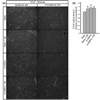
Protection of the nigral dopaminergic neurons and terminal fibers by enriched environment (EE). (a) Microphotographs of rat midbrain sections stained for tyrosine hydroxylase (TH) immunohistochemistry 21 days post-lesion. (b) Dopaminergic neuron survival in the substantia nigra 21 days after the 6-OHDA injection (lesion) in rats housed in EE (6-OHDA + EE) or in standard condition (6-OHDA). Panel (c) exhibits the mean value (six sections per animal) for each of the animals of panel (b). Panel (d) shows photomicrographs of rat striatal sections stained with TH 21 days following 6-OHDA injection after EE (6-OHDA + EE) or standard housing (6-OHDA). Bars represent mean ± SEM. Sham, n = 3; Sham + EE, n = 3; 6-OHDA, n = 6; 6-OHDA + EE, n = 11 (*p < 0.05). TH-IR: tyrosine hydroxylase immunoreactive. Panel (a) scale bar: 300 µm. Panel (d) scale bar: 0.5 mm.
Fig. 2
Fig. 2
Structural protection of the nigrostriatal pathway by enriched environment (EE). (a) Microphotographs of rat midbrain sections stained for FG immunohistochemistry 21 days post-lesion. (b) Percentage of FG-stained neurons in the substantia nigra of rats 21 days after 6-OHDA and housed in EE (6-OHDA + EE) or standard conditions (6-OHDA). Bars represent mean ± SEM, n = 5 per group (*p < 0.05). FG-IR, Fluorogold immunoreactive. Scale bar: 300 µm.
Fig. 3
Fig. 3
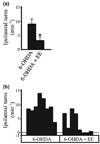
Functional protection of the nigrostriatal system by enriched environment (EE). (a) Results of amphetamine-induced rotation test 21 days after 6-OHDA injection. (b) Rotation behavior of individual rats. Experimental groups: 6-OHDA (toxin injected and standard housing) and 6-OHDA + EE (toxin injected and EE housing). Bars represent mean ± SEM, n = 8 per group (*p < 0.05).
Fig. 4
Fig. 4
Enriched environment (EE) prevents early neurodegeneration induced by 6-OHDA. (a) Representative brain coronal section drawing showing an arbitrary division in anterior and posterior substantia nigra (SN). (b) Rat midbrain sections of the ipsilateral anterior SN stained with the neurodegeneration marker Fluoro Jade-B (FJB). (c) Table showing anterior nigral neurodegeneration in contrast with an unstained posterior SN. Anterior values are the mean FJB+ cell number from the −4.52, −4.8, and −5.2 mm sections from bregma (±SEM); posterior values are the mean FJB+ cell number from the −5.6, −6.04, and −6.3 mm sections from bregma (±SEM). Experimental groups: sham (vehicle injected and standard housing), sham + EE (vehicle injected and EE housing), 6-OHDA (toxin injected and standard housing), and 6-OHDA + EE (toxin injected and EE housing) animals. n = 5 per group (*p < 0.05). Scale bar: 150 µm.
Fig. 5
Fig. 5
Enriched environment (EE) does not prevent 6-OHDA-induced loss of tyrosine hydroxylase (TH) 4 days after the injection. (a) TH cells 4 days after the 6-OHDA administration in sham (vehicle injected and standard housing), sham + EE (vehicle injected and EE housing), 6-OHDA (toxin injected and standard housing), and 6-OHDA + EE (toxin injected and EE housing) animals. Number of FJB+ cells was obtained by averaging three sections for the anterior SNpc and three sections for the posterior SNpc. (b) 6-OHDA administration did not decrease FG-labeled neurons 4 days after the injection. (c) Striatal TH levels 4 days after 6-OHDA. (d) Microphotographs of rat anterior midbrain sections stained for TH and FG immunohistochemistry 4 days after the toxin injection. Bars represent mean ± SEM. n = 5 per group (*p < 0.05). TH-IR, tyrosine hydroxylase immunoreactive; FGIR, Fluorogold immunoreactive. Scale bar: 100 µm.
Fig. 6
Fig. 6
Astrocytic reaction in the substantia nigra pars compacta (SNpc) 4 days after 6-OHDA. (a) Microphotographs of rat midbrain sections stained for glial fibrillary acidic protein (GFAP) immunohistochemistry 4 days after 6-OHDA injection. Experimental groups: 6-OHDA (toxin injected and standard condition housing) and 6-OHDA + EE (toxin injected and EE housing) animals. White line was drawn using TH immunostaining of the same section as reference, and limits the SNpc area. (b) GFAP optical density quantification 4 days post-toxin administration. Determinations were assessed in the anterior and posterior. Experimental groups: sham (vehicle injected and standard condition housing), sham + EE (vehicle injected and EE housing), 6-OHDA (toxin injected and standard housing), and 6-OHDA + EE (toxin injected and EE housing). Bars represent mean ± SEM. n = 5 per group (*p < 0.05). Scale bar: 300 µm.
Fig. 7
Fig. 7
Striatal astrocytic reaction 4 days after 6-OHDA. (a) Microphotographs of rat striatal sections stained for glial fibrillary acidic protein (GFAP) immunohistochemistry 4 days after the injection. Experimental groups: sham (vehicle injected and standard housing), sham + EE (vehicle injected and EE housing), 6-OHDA (toxin injected and standard housing), and 6-OHDA + EE (toxin injected and EE housing) animals. (b) GFAP optical density quantification 4 days post-toxin administration. Bars represent mean ± SEM. n = 5 per group (*p < 0.05). Scale bar: 300 µm.

References

    1. Anastasía A, De Erausquin GA, Wojnacki J, Mascó DH. Protection of dopaminergic neurons by electroconvulsive shock in an animal model of Parkinson’s disease. J. Neurochem. 2007;103:1542–1552. - PubMed
    1. Aponso P, Faull R, Connor B. Increased progenitor cell proliferation and astrogenesis in the partial progressive 6-hydroxydopamine model of Parkinson’s disease. Neuroscience. 2008;151:1142–1153. - PubMed
    1. Bezard E, Dovero S, Belin D, Duconger S, Jackson-Lewis V, Przedborski S, Piazza PV, Gross CE, Jaber M. Enriched environment confers resistance to 1-methyl-4-phenyl-1,2,3,6-tetrahydropyridine and cocaine: involvement of dopamine transporter and trophic factors. J. Neurosci. 2003;23:10999–11007. - PMC - PubMed
    1. Bowenkamp K, David D, Lapehak P, Henry M, Granholm A, Hoffer BJ, Mahalik T. 6-Hydroxydopamine induces the loss of the dopaminergic phenotype in substantia nigra neurons of the rat. Exp. Brain Res. 1996;111:7–9. - PubMed
    1. Carman LS, Gage FH, Shults CW. Partial lesion of the substantia nigra: relation between extent of lesion and rotational behaviour. Brain Res. 1991;553:275–283. - PubMed

Publication types

MeSH terms