

Gene expression markers of tendon fibroblasts in normal and diseased tissue compared to monolayer and three dimensional culture systems
- PMID: 19245707
- PMCID: PMC2651848
- DOI: 10.1186/1471-2474-10-27
Gene expression markers of tendon fibroblasts in normal and diseased tissue compared to monolayer and three dimensional culture systems
Abstract
Background: There is a paucity of data regarding molecular markers that identify the phenotype of the tendon cell. This study aims to quantify gene expression markers that distinguish between tendon fibroblasts and other mesenchymal cells which may be used to investigate tenogenesis.
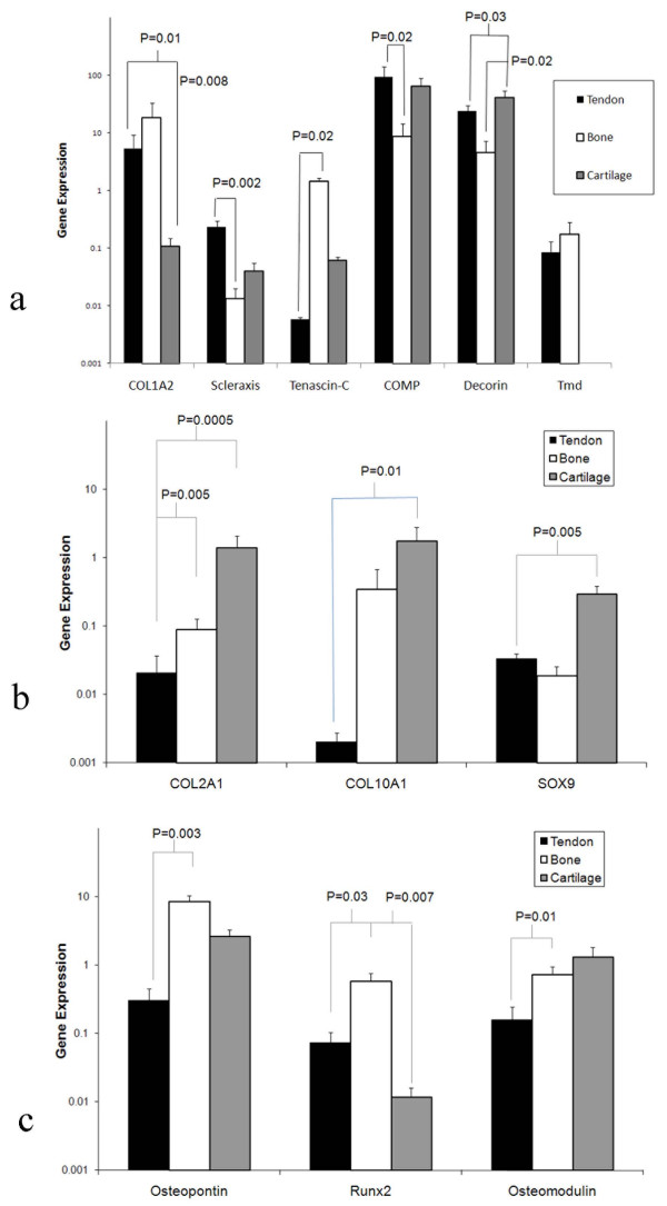
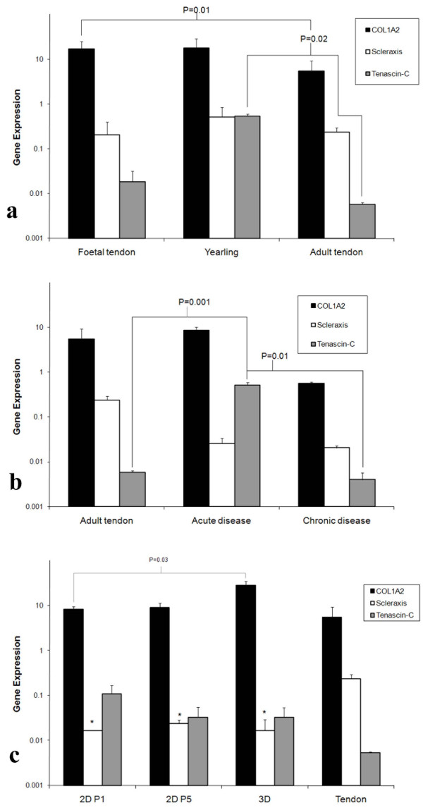
Methods: Expression levels for 12 genes representative of musculoskeletal tissues, including the proposed tendon progenitor marker scleraxis, relative to validated reference genes, were evaluated in matched samples of equine tendon (harvested from the superficial digital flexor tendon), cartilage and bone using quantitative PCR (qPCR). Expression levels of genes associated with tendon phenotype were then evaluated in healthy, including developmental, and diseased equine tendon tissue and in tendon fibroblasts maintained in both monolayer culture and in three dimensional (3D) collagen gels.
Results: Significantly increased expression of scleraxis was found in tendon compared with bone (P = 0.002) but not compared to cartilage. High levels of COL1A2 and scleraxis and low levels of tenascin-C were found to be most representative of adult tensional tendon phenotype. While, relative expression of scleraxis in developing mid-gestational tendon or in acute or chronically diseased tendon did not differ significantly from normal adult tendon, tenascin-C message was significantly upregulated in acutely injured equine tendon (P = 0.001). Relative scleraxis gene expression levels in tendon cell monolayer and 3D cultures were significantly lower than in normal adult tendon (P = 0.002, P = 0.02 respectively).
Conclusion: The findings of this study indicate that high expression of both COL1A2 and scleraxis, and low expression of tenascin-C is representative of a tensional tendon phenotype. The in vitro culture methods used in these experiments however, may not recapitulate the phenotype of normal tensional tendon fibroblasts in tissues as evidenced by gene expression.
Figures
References
-
- Patterson-Kane JC, Firth EC. The pathobiology of exercise-induced superficial digital flexor tendon injury in Thoroughbred racehorses. Vet J. 2008. - PubMed
Publication types
MeSH terms
Substances
LinkOut - more resources
Full Text Sources
Other Literature Sources
Medical
Miscellaneous
