. 2009 Apr 21;106(16):6471-6.
doi: 10.1073/pnas.0810034106. Epub 2009 Feb 25.

Exploring the roles of noise in the eukaryotic cell cycle

Affiliations

Exploring the roles of noise in the eukaryotic cell cycle

Sandip Kar et al. Proc Natl Acad Sci U S A. .

Abstract

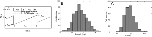
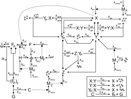
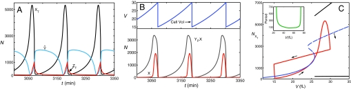
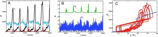
The DNA replication-division cycle of eukaryotic cells is controlled by a complex network of regulatory proteins, called cyclin-dependent kinases, and their activators and inhibitors. Although comprehensive and accurate deterministic models of the control system are available for yeast cells, reliable stochastic simulations have not been carried out because the full reaction network has yet to be expressed in terms of elementary reaction steps. As a first step in this direction, we present a simplified version of the control system that is suitable for exact stochastic simulation of intrinsic noise caused by molecular fluctuations and extrinsic noise because of unequal division. The model is consistent with many characteristic features of noisy cell cycle progression in yeast populations, including the observation that mRNAs are present in very low abundance (approximately 1 mRNA molecule per cell for each expressed gene). For the control system to operate reliably at such low mRNA levels, some specific mRNAs in our model must have very short half-lives (<1 min). If these mRNA molecules are longer-lived (perhaps 2 min), then the intrinsic noise in our simulations is too large, and there must be some additional noise suppression mechanisms at work in cells.


Conflict of interest statement

The authors declare no conflict of interest.

Figures

Fig. 1.
Fig. 1.
Cell cycle statistics. (A) Schematic illustration of a cell lineage undergoing growth and division. Each cell is born at a particular size (Sbir) and divides at some larger size (Sdiv). The birth size of a daughter cell is some fraction p of the division size of its mother, where p is a random number drawn from a normal distribution with mean 0.5 and 5% CV. The cell cycle time (Tcc) is the time between cell birth and division; it may also be called cell age at division. The interdivision period is divided into four phases, depending on the state of the chromosomal DNA: G1 (unreplicated chromosomes), S (replicating chromosomes), G2 (replicated chromosomes), M (dividing chromosomes). During G1 phase, CDK activity is low, whereas CDK activity is high during S + G2 + M phases. (B and C) Histograms for cell length (B) and age (C) in a sample of dividing fission yeast cells. Data were redrawn from Miyata et al. (23). Fission yeast cells are rod-shaped; they grow in length only at a fixed radius, so cell length is a proxy for cell size. For this yeast cell sample, mean size at division = 13.4 μm (CV = 7.5%), and mean age at division = 116 min (CV = 13.8%).
Fig. 2.
Fig. 2.
Molecular mechanism regulating the activity of cyclin B-dependent protein kinase. X, CycB–Cdk1; Y, Cdh1—APC; YP, phosphorylated (inactive) Y; Z, Cdc20 and Cdc14 (composite species); G, gene encoding Z; F, transcription factor controlling the expression of G; FP, phosphorylated form of F; H, enzyme that removes phosphate group from FP; (FP)2, dimeric form of F; C, Z gene bound to (FP)2 and actively transcribing MZ, the messenger RNA for Z; MX etc., messenger RNAs for all other primary gene products in the model; four small circles, products of protein and mRNA degradation reactions. A T-shaped arrow with balls on the cross-bar indicates a reversible binding reaction. (Inset) Additional degradation reactions necessary to maintain approximately constant concentrations of total Y and F proteins during the cell cycle. We assume that the complex YP–X has enough CDK activity to drive DNA synthesis but not mitosis; only the free form of X is sufficient to drive the cell into mitosis.
Fig. 3.
Fig. 3.
Deterministic simulations of the model. (A) Time courses for total amounts of cyclin B protein (XT, black line), Cdc20 protein (ZT, red line), and unphosphorylated Cdh1 protein (Ŷ blue line). (B) Time courses for free (uncomplexed) X (red line), YP–X (gray line), and cell volume (blue line). (C) The figure 8-shaped curve (red) is a cell cycle trajectory created by plotting, parametrically in t, the curves for XT(t) and V(t) from A and B, respectively. The small arrows indicate the direction of motion around this trajectory. Cell division is indicated by the abrupt jump from V = 29.8 to V = 14.9, which is triggered when Ŷ increases above 1,200 molecules. The S-shaped curve (blue) is a one-parameter bifurcation diagram (computed by using XPP-AUTO) for steady-state solutions of the molecular control system, treating cell size (V) as a bifurcation parameter. Solid line, stable steady state; dashed line, unstable steady state. The two black lines above and below the unstable steady-state curve indicate the presence of stable limit cycle oscillations for V > 25; the Upper (Lower) curve indicating the maximum (minimum) value of XT on the limit cycle for a particular value of V. (Inset) Period of limit cycle oscillations as a function of cell size. The control system generates oscillations by homoclinic (infinite period) bifurcations at V = 25 and V = 74.
Fig. 4.
Fig. 4.
Stochastic simulations of the full model. These simulations include both intrinsic noise (fluctuations in the molecular regulatory system) and extrinsic noise (uneven cell division and binomial distribution of protein and mRNA molecules to daughter cells at division). (A) Time courses for total amounts of regulatory proteins, as in Fig. 3A. (B) Time courses for amounts of mRNA species, MZ (Upper) and MX (Lower). As described in the text, we assume that MZ has a half-life of 280 s, whereas MX (as well as all other mRNAs) has a much shorter half-life (12 s). (C) Series of stochastic cell cycle trajectories are superimposed on the bifurcation diagram of Fig. 3C.

References

    1. Tyson JJ, Novak B. Temporal organization of the cell cycle. Curr Biol. 2008;18:R759–R768. - PMC - PubMed
    1. Tyson JJ. The coordination of cell growth and division: Intentional or incidental? Bioessays. 1985;2:72–77.
    1. Fantes PA. Control of cell size and cycle time in Schizosaccharomyces pombe. J Cell Sci. 1977;24:51–67. - PubMed
    1. Koch AL, Schaechter M. A model for statistics of the cell division process. J Gen Microbiol. 1962;29:435–454. - PubMed
    1. Swain PS, Elowitz MB, Siggia ED. Intrinsic and extrinsic contributions to stochasticity in gene expression. Proc Natl Acad Sci USA. 2002;99:12795–12800. - PMC - PubMed

Publication types

MeSH terms

Substances

LinkOut - more resources