. 2009 May 1;284(18):12432-46.
doi: 10.1074/jbc.M806772200. Epub 2009 Feb 26.

Delivery of CD44 shRNA/nanoparticles within cancer cells: perturbation of hyaluronan/CD44v6 interactions and reduction in adenoma growth in Apc Min/+ MICE

Affiliations

Delivery of CD44 shRNA/nanoparticles within cancer cells: perturbation of hyaluronan/CD44v6 interactions and reduction in adenoma growth in Apc Min/+ MICE

Suniti Misra et al. J Biol Chem. .

Abstract

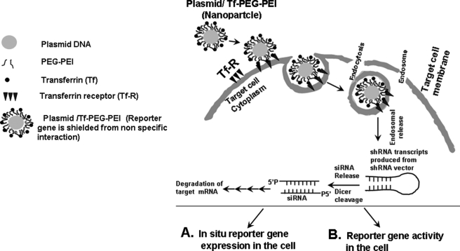
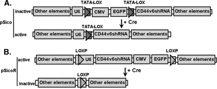
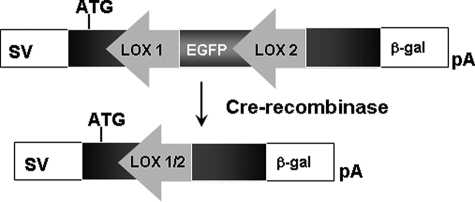
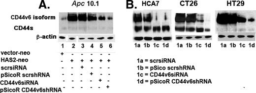
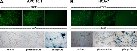
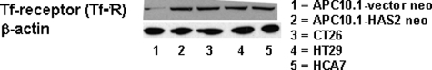
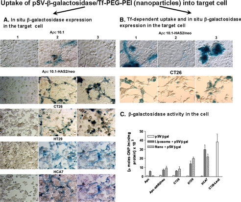
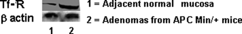
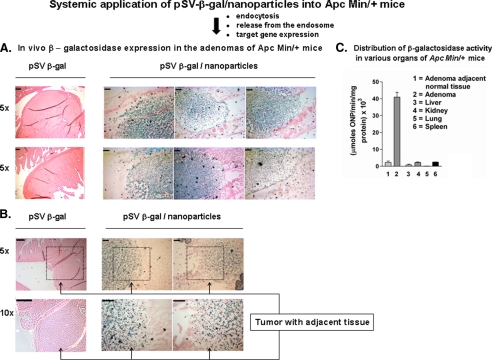
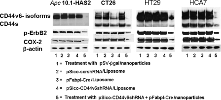
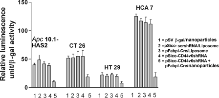
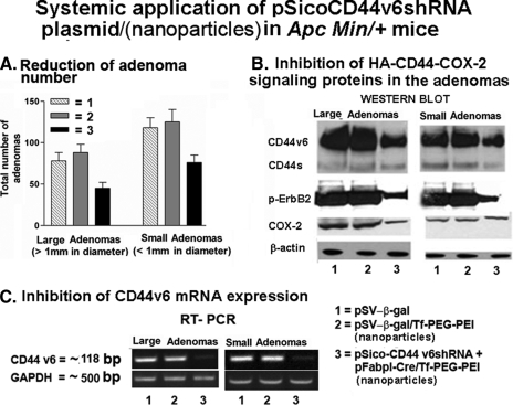
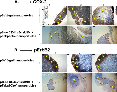
Our studies have shown that constitutive interactions between hyaluronan and CD44 on tumor cells induces various anti-apoptotic cell survival pathways through the formation of a multimeric signaling complex that contains activated receptor tyrosine kinases. Inhibition of the hyaluronan-CD44 interactions on tumor cells by hyaluronan-CD44 interaction antagonists suppresses these activities by disassembling the complex. Although the anti-tumor activity of hyaluronan-oligosaccharides, a hyaluronan-CD44 interaction antagonist, is effective in sensitizing tumor cells to chemotherapeutic agents and reducing tumor growth in xenografts, hyaluronan-oligosaccharide alone was not effective in reducing tumor progression in Apc Min/+ mice. We now show in vitro and in vivo that targeted inhibition of the expression of CD44v6 depletes the ability of the colon tumor cells to signal through hyaluronan-CD44v6 interactions. First, we cloned oligonucleotides coding CD44v6 shRNA into a conditionally silenced pSico vector. Second, using pSico-CD44v6 shRNA and a colon-specific Fabpl promoter-driven Cre recombinase expression vector packaged into transferrin-coated nanoparticles, we successfully delivered the CD44v6 shRNA within pre-neoplastic and neoplastic colon malignant cells. Third, using the Apc Min/+ mice model, we demonstrated that inhibition of the CD44v6 expression reduces the signaling through a hyaluronan/CD44v6-pErbB2-Cox-2 interaction pathway and reduced adenoma number and growth. Together, these data provide insight into the novel therapeutic strategies of short hairpin RNA/nanoparticle technology and its potential for silencing genes associated with colon tumor cells.


Figures

FIGURE 1.
FIGURE 1.
Schematic illustration of cellular uptake of plasmid DNA/Tf-PEG-PEI (nanoparticles) polyplexes, their shielding from nonspecific interaction, and mechanism of action of shRNA. Internalization of PEG-shielded and Tf-R-targeted polyplexes into target cells occurs by receptor-mediated endocytosis after association of polyplex ligand Tf to Tf-R present on the target cell plasma membrane. Internalized particles are trafficked to endosomes followed by endosomal release of the particles and/or the nucleic acid into cytoplasm. Released siRNA will be induced to RNA-induced silencing complex and will be guided for cleavage of complementary target mRNA in the cytoplasm. siRNA (antisense) guide strand will direct the targeted RNAs to be cleaved by RNA endonuclease. Finally plasmid/Tf-PEG-PEI-nanoparticles delivery in the target cell shows reporter gene expression and activity.
FIGURE 2.
FIGURE 2.
Description of pSico and pSicoR vectors (53). A, section of pSico vector. In pSico, the U6 promoter that drives the expression of shRNA is made conditional in two steps. First, bifunctional TATAlox boxes are placed in place of TATA box sites so that the resulting promoter retains transcriptional activity. Second, a cytomegalovirus (CMV)-enhanced EGFP stop/reporter cassette was inserted between two TATALox sites so that after Cre-mediated recombination the CMV-EGFP cassette will be excised to generate a functional U6 promoter. A T6 sequence is placed immediately upstream of U6 and serves as a stop signal of U6 promoter. Oligonucleotides coding shRNA are cloned into HpaI-XhoI-digested pSico vectors. Cre recombinase eliminates the CMV-EGFP cassette. Transcription of CD44v6 shRNA from the U6 promoter will ensue. B, section of pSicoR vector. In pSicoR the CMV-EGFP cassette is placed downstream of the U6 promoter and does not affect its activity. Two LoxP sites in the same orientation are present in this vector. Oligonucleotides coding CD44v6 shRNA are cloned into HpaI-XhoI-digested pSicoR vectors. The transcription from U6 promoter will produce CD44v6 shRNA constitutively in the absence of Cre recombinase. In the presence of Cre recombinase, the CD44v6 shRNA production unit and CMV-EGFP cassette are eliminated.
FIGURE 3.
FIGURE 3.
Validity of tissue-specific promoter using a gene-switch reporter construct. The specificity of promoter-driven Cre recombinase was determined by using a gene-switch reporter construct. The construct is an SV40 promoter-driven gene-switch plasmid containing floxed EGFP inserted into the open reading frame of the β-galactosidase (β-gal) gene resulting in appropriate translation of the EGFP gene showing green fluorescence but not the β-galactosidase gene. In the presence of appropriate Cre recombinase, the EGFP gene is excised, and the correct reading frame of the β-galactosidase gene is restored, which is now expressed, and blue color in Fig. 5 indicates that it can react with X-gal to produce blue dye.
FIGURE 4.
FIGURE 4.
Inhibition of CD44 variants by CD44v6 siRNA and CD44v6 shRNA. A, Apc 10.1 cells were transfected with vector-neo or HAS2-neo and co-transfected with control scrsiRNA, or control pSicoR-scrshRNA, or CD44v6 siRNA, or pSicoR-CD44v6 shRNA plasmids. Transient transfection was carried out at 100 pmol of siRNA using Oligofectamine or 0.5 μg of shRNA using Lipofectamine (Invitrogen) according to the manufacturer's instructions. Cells were transfected with the siRNA or shRNA in 6-well plates with cells at 70-90% confluence. The cells were then incubated at 37 °C in 5% CO2 for 24 h, replated in 150-mm dishes, and allowed to grow for 48 h in complete medium as described under “Experimental Procedures.” B, similarly, HCA7, CT26, and HT29 colon cancer cells were transfected with the plasmids 1a-1d as indicated. The transfectants were grown for 72 h after transfections. Western blots of cell lysates were done with human CD44 and β-actin (loading control) antibodies.
FIGURE 5.
FIGURE 5.
Tissue-specific gene expression with a gene-switch reporter construct. Transient transfection was carried out at 0.5 μg of plasmid using Lipofectamine (Invitrogen) according to the manufacturer's instructions. Cells were transfected with the plasmid in 6-well plates with cells at 70-90% confluence as described under “Experimental Procedures.” The cells were then incubated at 37 °C in 5% CO2 for 24 h, replated in 150-mm dishes, and allowed to grow for 48 h in complete medium. Apc 10.1 and HCA7 cells transfected with the pSV-EGFP-β-gal show EGFP expression in most, if not all, cells (upper left panels of A and B). Co-transfection with the nonspecific promoter-driven Cre cDNA plasmid (pProbasin-Cre) shows the same result (upper middle panels of A and B). Co-transfection with the tissue-specific promoter-driven Cre plasmid (pFabpl-Cre) eliminates the EGFP expression (upper right panels of A and B) with concomitant expression of β-galactosidase as demonstrated by X-gal staining (lower right panels of A and B). Cultures without the tissue-specific promoter did not express β-galactosidase (lower left and middle panels of A and B).
FIGURE 6.
FIGURE 6.
Elevated expression of HAS2 stimulates expression of Tf-R protein in the colon pre-neoplastic (Apc 10. 1) cells and is present at high levels in the cancer cells. Apc 10.1 cells were transfected with the vector-neo (lane 1) or HAS2-neo cDNA (lane 2). These transfectants as well as HCA7, CT26, and HT29 colon cancer cells were grown for 72 h, and Western blots of cell lysates were analyzed for transferrin receptor and β-actin as a loading control. The data presented in this figure are representative of three independent experiments.
FIGURE 7.
FIGURE 7.
Delivery of pSV-β-galactosidase-nanoparticles in cell culture. A, in situ β-galactosidase expression in the target cell. The Apc 10.1 cells, Apc 10.1-HAS2 clone, and the CT26, HT29, and HCA7 cells were treated for 48 h with the pSV-β-gal alone (panel 1), or with the pSV-β-gal with liposome (panel 2), or with the pSV-β-gal/nanoparticles (35 nm average diameter, 8 μg of pSV-β-gal/ml) (panel 3). The average size of the pSV-β-gal/nanoparticles is ∼35 nm ± 20 nm (Table 1). The transfected cells were fixed in 0.2% glutaraldehyde in PBS and washed twice in PBS as described under “Experimental Procedures.” The cells were treated with a β-galactosidase staining solution as described under “Experimental Procedures ” and digitally photographed. B, transferrin-dependent uptake and in situ β-galactosidase expression in the target cell. The Apc 10.1-HAS2 cells were transfected with the pSV-β-gal with liposome (panel 1), or treated with Tf-R antibody and followed by transfection with the pSV-β-gal/nanoparticles (8 μg pSV-β-gal/ml) (panel 2), or treated with the pSV-β-gal/nanoparticles (8 μg pSV-β-gal/ml) alone (panel 3). The transfected cells were fixed in 0.2% glutaraldehyde in PBS and washed twice in PBS as described under “Experimental Procedures.” The cells were treated with a β-galactosidase staining solution as described under “Experimental Procedures ” and digitally photographed. C, cell-free extracts of parallel cultures were prepared in 10 mm CHAPS buffer and assayed for β-galactosidase activity using o-nitrophenyl β-d-galactopyranoside as substrate as described under “Experimental Procedures.” The results are expressed as micromoles of o-nitrophenol formed per min/mg protein and represent ± S.D. of triplicate assays from the untransfected, or liposome-transfected, or nanoparticles-treated cultures for each cell type.
FIGURE 8.
FIGURE 8.
Elevated expression of Tf-R in Apc Min/+ mice adenomas compared with adjacent normal colon mucosa. The tissue extracts from adenomas and adjacent normal tissue were processed for Western blot and immunoblotted with Tf-R antibody and β-actin (as loading control). The data presented in this figure are representative of three independent experiments.
FIGURE 9.
FIGURE 9.
Delivery of pSV-β-gal nanoparticles in Apc Min/+ mice. A, in vivo β-galactosidase expression in the adenomas of Apc Min/+ mice. Ten Apc Min/+ mice received pSV-β-gal plasmids (100 μg/100 μl, intraperitoneally) alone and 10 received pSV-β-galactosidase (pSV-β-gal)/nanoparticles (100 μg/100 μl, intraperitoneally) every other day. Mice showed no sign of toxicity throughout the experiment. At day 10, mice were euthanized with isoflurane in a vented chemical hood followed by cervical dislocation. Livers, spleens, kidneys, and lungs were harvested and frozen immediately in dry ice as described under “Experimental Procedures.” After counting the tumors, the flattened intestines from various treatment groups such as pSV-β-gal (group 1), pSV-β-gal/nanoparticles (group 2) were rolled into Swiss rolls and fixed in OCT on dry ice. Frozen sections carrying the adenomas were stained with X-gal (blue dye deposits around the tumor tissue at ×5 magnification) and eosin, whereas the tumor bearing animals treated with pSV-β-gal showing no lacZ stain were outlined with a black line. To visualize whether low lacZ expression occurs in the surrounding tissue, the sections were examined at low (×5) and high (×10) magnifications. Scale bars, 200 μm (B). The selected portions in black square area at ×5 magnification are shown at ×10 magnification. The normal mucosal epithelium (at ×10 magnification) shows little or no lacZ expression (B). Scale bars, 200 μm. C, β-galactosidase activity in adenomas, adjacent normal tissue and other organs of Apc Min/+ mice. From the second part of the groups 1-3, mucosa and enterocytes were removed from segments of intestine by lightly scraping the mucosal surface with the edge of a microscope slide, and tumors were excised and pooled. The tumor and adjacent normal tissues were subsequently used for measuring β-galactosidase activity as described under “Experimental Procedures.” The enzyme assays in the tissue extracts were done in three sets of individual experiments. The graph shows a representative result (average of n = 3 ± S.D.).
FIGURE 10.
FIGURE 10.
(pSico-CD44v6 shRNA plus pFabpl-Cre)/nanoparticles knock down CD44v6, COX-2, and p-ErbB2 in colon pre-neoplastic and cancer cells. Apc 10.1-HAS2 clone cells and CT26, HT29, and HCA7 colon cancer cells were transfected with liposomes and either pSico-scrshRNA, or pFabpl-Cre, or pSico-CD44v6 shRNA, or were treated with nanoparticles containing either pSV-β-gal or the combination of pSico-CD44v6 shRNA plus pFabpl-Cre and cultured for 72 h. Lysates were prepared and probed in Western blots with antibodies to COX-2, human CD44, p-ErbB2, and β-actin. Total ErbB2 remained unchanged in all the treatment groups (data not shown).
FIGURE 11.
FIGURE 11.
(pSico-CD44v6 shRNA plus pFabpl-Cre)/nanoparticles suppress COX-2 reporter gene expression in vitro. Apc 10.1-HAS2 clone cells and CT26, HT29, and HCA7 colon cancer cells were co-transfected for 12 h with the pCOX-2 luciferase and the pSV-β-gal reporter plasmids. They were then further transfected for 72 h with liposomes plus either pSico-scrshRNA, or pFabpl-Cre, or pSico-CD44v6 shRNA, or were treated with nanoparticles that contained either pSV-β-gal or the combined pSico-CD44v6 shRNA plus pFabpl-Cre. Cells were extracted with reporter lysis buffer followed by measurement of luciferase and β-galactosidase activity. The graph shows one of three experiments with nearly equivalent results and gives the average ± S.D. of three measurements for each culture.
FIGURE 12.
FIGURE 12.
Reduction in number of adenomas, protein expression, and RT-PCR analysis in Apc Min/+ mice adenomas after 10 days of plasmid-nanoparticles treatment. A, inhibition of adenoma growth. Thirty Apc Min/+ mice were randomly divided into three groups. Group 1 received pSV-β-galactosidase (100 μg/100 μl, intraperitoneally) alone. Group 2 received pSV-β-gal nanoparticles (100 μg/100 μl, intraperitoneally) targeted to the Tf-R. Group 3 received pSico-CD44v6 shRNA (75 μg) plus pFabpl-Cre (25 μg)/nanoparticles intraperitoneally every other day. 10 days after the beginning of treatment (i.e. 1 day after the last injection of the plasmid), the animals were sacrificed, and the large (>1 mm) and small (<1 mm) adenomas were counted. Large and small adenomas were excised 10 days after the pSico-CD44v6 shRNA plus pFabpl-Cre-/nanoparticles treatment. The control experiment with pSico-CD44v6 shRNA/nanoparticles or pFabpl-Cre/nanoparticles gave similar results as pSV-β-gal/nanoparticles as described under “Experimental Procedures.” After counting the tumors in groups 1-3, mucosa and enterocytes were removed from segments of intestine by lightly scraping the mucosal surface with the edge of a microscope slide and tumors were excised and pooled. The tumor and adjacent normal tissues were subsequently used for Western blot and RT-PCR analysis. B, Western blot analysis. Tissue extracts were processed for Western blot analysis of CD44, pErbB2, TErbB2, Cox-2, and β-actin. Total ErbB2 remained unchanged in all the treatment groups (data not shown). C, RT-PCR. Total RNAs were extracted from the above set of experiments and analyzed by RT-PCR for mouse CD44v6 and glyceraldehyde-3-phosphate dehydrogenase (GAPDH) expression.
FIGURE 13.
FIGURE 13.
Analysis of Cox-2 and pErbB2 in the paraffin sections of intestines having tumor tissues from various treatment groups of three different Apc Min/+ mice were demonstrated from the experiment as described in Fig. 12 legend. Panels 1-3 refer to different animals. For immunohistochemical analysis, antigens were retrieved from tissue sections from treated and untreated tumors and then treated with antibodies against Cox-2 (A) and pErbB2 (B) following standard avidin-biotin-peroxidase complex method. After probing for pErbB2 and Cox-2, the sections were counter-stained with hematoxylin. The size-matched tumors (outlined with red dotted lines) from various treatments were compared. The upper panels in each case are adenomas from mice treated with pSV-β-gal nanoparticles (controls) that stain heavily for Cox-2 and p-ErbB2 (HRP stain, indicated by yellow arrows) as expected from their high expression in these cells. In contrast, sections in the lower panels in each case are adenomas from mice treated with the nanoparticles that contain pSico-CD44v6 shRNA plus pFabpl-Cre plasmids show smaller sparse or negligible HRP staining for Cox-2 and pErbB2. Importantly, it should be noted that the normal intestine villi treated with (pSico-CD44v6 shRNA plus pFabpl-Cre)/nanoparticles stained heavily for Cox-2 and pErbB2, demonstrating the specificity of the nanoparticle for the adenoma (to target CD44v6 mRNA) versus normal intestine. CD44v6 shRNA plus pFabpl-Cre plasmid/nanoparticle treatment did not show any significant change in the HA content in tumor sections (data not shown). Scale bars, 200 μm.

References

    1. Sprenger, C. C., Plymate, S. R., and Reed, M. J. (2008) Br. J. Cancer 98 250-255 - PMC - PubMed
    1. Markwald, R. R., Fitzharris, T. P., Bank, H., and Bernanke, D. H. (1978) Dev. Biol. 62 292-316 - PubMed
    1. Lee, J. Y., and Spicer, A. P. (2000) Curr. Opin. Cell Biol. 12 581-586 - PubMed
    1. Toole, B. P. (2004) Nat. Rev. Cancer 4 528-539 - PubMed
    1. Hascall, V. C., Majors, A. K., De La Motte, C. A., Evanko, S. P., Wang, A., Drazba, J. A., Strong, S. A., and Wight, T. N. (2004) Biochim. Biophys. Acta 1673 3-12 - PubMed

Publication types

MeSH terms