

Brain nuclear factor-kappa B activation contributes to neurohumoral excitation in angiotensin II-induced hypertension
- PMID: 19246475
- PMCID: PMC2682616
- DOI: 10.1093/cvr/cvp073
Brain nuclear factor-kappa B activation contributes to neurohumoral excitation in angiotensin II-induced hypertension
Abstract
Aims: Angiotensin II (ANG II)-induced inflammatory and oxidative stress responses contribute to the pathogenesis of hypertension. In this study, we determined whether nuclear factor-kappa B (NF-kappaB) activation in the hypothalamic paraventricular nucleus (PVN) increases oxidative stress and contributes to the ANG II-induced hypertensive response.
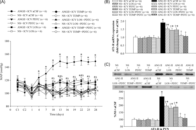
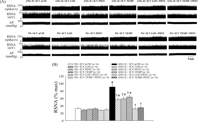
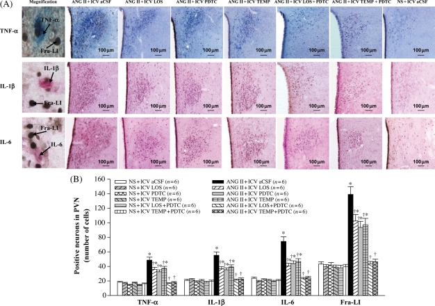
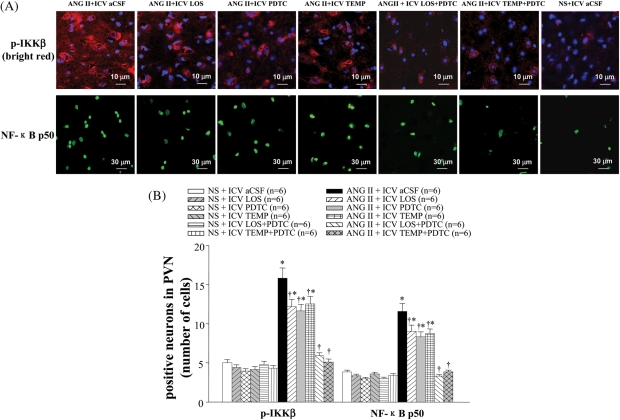
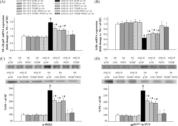
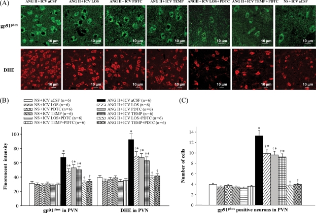
Methods and results: Rats were infused intravenously with ANG II (10 ng/kg per min) or saline for 4 weeks. These rats received either vehicle or losartan (LOS, 20 microg/h), an angiotensin II type 1 receptor (AT1-R) antagonist; pyrrolidine dithiocarbamate (PDTC, 5 microg/h), a NF-kappaB inhibitor; tempol (TEMP, 80 microg/h), a superoxide scavenger; LOS (20 microg/h), and PDTC (5 microg/h); or TEMP (80 microg/h) and PDTC (5 microg/h), given intracerebroventricularly (ICV) via osmotic minipump. ANG II infusion resulted in increased mean arterial pressure, renal sympathetic nerve activity, plasma proinflammatory cytokines (PIC), norepinephrine, and aldosterone. These rats also had higher levels of Fra-LI (an indicator of chronic neuronal activation), PIC, phosphorylated IKKbeta, NF-kappaB subunits, AT1-R, superoxide, and gp91phox (a subunit of NADP(H) oxidase) and lower levels of IkappaBalpha in the PVN than control animals. ICV treatment with LOS, PDTC, or TEMP attenuated these changes, and combined treatment with ICV LOS and PDTC, or ICV TEMP and PDTC prevented these ANG II-induced hypertensive responses.
Conclusion: These findings suggest that an ANG II-induced increase in the brain renin-angiotensin system activates NF-kappaB in the PVN and contributes to sympathoexcitation in hypertension. The increased superoxide in the PVN contributes to NF-kappaB activation and neurohumoral excitation in hypertension.
Figures

Similar articles
-
Cross-talk between cytokines and renin-angiotensin in hypothalamic paraventricular nucleus in heart failure: role of nuclear factor-kappaB.Cardiovasc Res. 2008 Sep 1;79(4):671-8. doi: 10.1093/cvr/cvn119. Epub 2008 May 10. Cardiovasc Res. 2008. PMID: 18469338 Free PMC article.
-
Interaction between AT1 receptor and NF-κB in hypothalamic paraventricular nucleus contributes to oxidative stress and sympathoexcitation by modulating neurotransmitters in heart failure.Cardiovasc Toxicol. 2013 Dec;13(4):381-90. doi: 10.1007/s12012-013-9219-x. Cardiovasc Toxicol. 2013. PMID: 23877628
-
Inhibition of reactive oxygen species in hypothalamic paraventricular nucleus attenuates the renin-angiotensin system and proinflammatory cytokines in hypertension.Toxicol Appl Pharmacol. 2014 Apr 15;276(2):115-20. doi: 10.1016/j.taap.2014.02.002. Epub 2014 Feb 25. Toxicol Appl Pharmacol. 2014. PMID: 24576725
-
Hypothalamic paraventricular nucleus activation contributes to neurohumoral excitation in rats with heart failure.Regen Med Res. 2014 Jan 8;2(1):2. doi: 10.1186/2050-490X-2-2. eCollection 2014 Dec. Regen Med Res. 2014. PMID: 25984330 Free PMC article. Review.
-
Mechanisms of renal sympathetic activation in renovascular hypertension.Exp Physiol. 2015 Apr 20;100(5):496-501. doi: 10.1113/expphysiol.2014.079855. Exp Physiol. 2015. PMID: 25639235 Review.
Cited by
-
Exercise Training Attenuates Hypertension via Suppressing ROS/MAPK/NF-κB/AT-1R Pathway in the Hypothalamic Paraventricular Nucleus.Nutrients. 2022 Sep 24;14(19):3968. doi: 10.3390/nu14193968. Nutrients. 2022. PMID: 36235619 Free PMC article.
-
Membrane trafficking of NADPH oxidase p47(phox) in paraventricular hypothalamic neurons parallels local free radical production in angiotensin II slow-pressor hypertension.J Neurosci. 2013 Mar 6;33(10):4308-16. doi: 10.1523/JNEUROSCI.3061-12.2013. J Neurosci. 2013. PMID: 23467347 Free PMC article.
-
Intracerebroventricular tempol administration in older rats reduces oxidative stress in the hypothalamus but does not change STAT3 signalling or SIRT1/AMPK pathway.Appl Physiol Nutr Metab. 2017 Jan;42(1):59-67. doi: 10.1139/apnm-2016-0067. Epub 2016 Oct 6. Appl Physiol Nutr Metab. 2017. PMID: 28006433 Free PMC article.
-
Chronic estrogen exposure affects gene expression in the rostral ventrolateral medulla of young and aging rats: Possible role in hypertension.Brain Res. 2015 Nov 19;1627:134-42. doi: 10.1016/j.brainres.2015.09.007. Epub 2015 Sep 12. Brain Res. 2015. PMID: 26375620 Free PMC article.
-
Central Blockade of E-Prostanoid 3 Receptor Ameliorated Hypertension Partially by Attenuating Oxidative Stress and Inflammation in the Hypothalamic Paraventricular Nucleus of Spontaneously Hypertensive Rats.Cardiovasc Toxicol. 2021 Apr;21(4):286-300. doi: 10.1007/s12012-020-09619-w. Epub 2020 Nov 9. Cardiovasc Toxicol. 2021. PMID: 33165770
References
-
- Simpson JB. The circumventricular organs and the central actions of angiotensin. Neuroendocrinology. 1981;32:248–256. - PubMed
-
- Zhu GQ, Gao L, Patel KP, Zucker IH, Wang W. ANG II in the paraventricular nucleus potentiates the cardiac sympathetic afferent reflex in rats with heart failure. J Appl Physiol. 2004;97:1746–1754. - PubMed
-
- Granger JP. An emerging role for inflammatory cytokines in hypertension. Am J Physiol Heart Circ Physiol. 2006;290:H923–H924. - PubMed
Publication types
MeSH terms
Substances
Grants and funding
LinkOut - more resources
Full Text Sources
Medical
Research Materials
Miscellaneous
