Review
. 2009 Jul;23(7):2024-33.
doi: 10.1096/fj.08-125971. Epub 2009 Feb 26.

Structure, expression, and biological function of INSM1 transcription factor in neuroendocrine differentiation

Affiliations
Review

Structure, expression, and biological function of INSM1 transcription factor in neuroendocrine differentiation

Michael S Lan et al. FASEB J. 2009 Jul.

Abstract

Zinc-finger transcription factors are DNA-binding proteins that are implicated in many diverse biological functions. INSM1 (formerly IA-1) contains five zinc-finger motifs and functions as a transcription factor. INSM1 protein structure is highly conserved in homologues of different species. It is predominantly expressed in developing neuroendocrine tissues and the nervous system in mammals. INSM1 represents an important player in early embryonic neurogenesis. In pancreatic endocrine cell differentiation, Ngn3 first activates INSM1 and subsequently NeuroD/beta2. Conversely, INSM1 exerts a feedback mechanism to suppress NeuroD/beta2 and its own gene expression. INSM1 gene ablation in the mouse results in the impairment of pancreatic endocrine cell maturation. Further, deletion of INSM1 severely impairs catecholamine biosynthesis and secretion from the adrenal gland that results in early embryonic lethality. Genetically, INSM1 acts as a downstream factor of Mash 1 and Phox2b in the differentiation of the sympatho-adrenal lineage. In the developing neocortex, mouse embryos lacking INSM1 expression contain half the number of basal progenitors and show a reduction in cortical plate radial thickness. Cell signaling studies reveal that INSM1 contributes to the induction of cell cycle arrest/exit necessary to facilitate cellular differentiation. INSM1 is highly expressed in tumors of neuroendocrine origin. Hence, its promoter could serve as a tumor-specific promoter that drives a specific targeted cancer gene therapy for the treatment of neuroendocrine tumors. Taken together, all of these features of INSM1 strongly support its role as an important regulator during neuroendocrine differentiation.


Figures

Figure 1.
Figure 1.
INSM1 structure. A) INSM1 mRNA is encoded by an intronless gene expanding a 2838-bp sequence with a 1530-bp coding sequence. The 510-aa protein can be divided into an N-terminal (aa 1-250) and a C-terminal (aa 251-510) domain. The N-terminal domain contains two proline-rich regions (aa 43-58 and 183-205, blue shaded area), several dibasic amino acids (green), and an amidation signal sequence (pink). The C-terminal domain contains five equally spaced zinc-finger motifs (red). B) Zinc-finger motifs of INSM1 were aligned with zinc-finger motifs derived from INSM1 homologues from other species. Cys2-His2 zinc fingers are highlighted in blue and pink. The first zinc-finger motif contains an Arg substitution for the last His residue in all species except D. melangaster, which has a Cys replacement (labeled with asterisk). C. elegans contains a perfect consensus zinc-finger 2, but the first and third fingers have either Arg or Gln substitutions for the last His residue. All of the 27 aa in the second zinc-finger motif among the homologues show a 96% identity.
Figure 2.
Figure 2.
Evolutionary conservation of INSM1 homologues. A) Schematic diagram of INSM1 homologues from human, chimpanzee, mouse, rat, X. laevis, zebrafish, D. melangaster, and C. elegans. Zinc-finger region represents a typical Cys2-His2-type zinc-finger motif. First zinc finger, however, is imperfect, and last His residue is replaced with an Arg or Cys (shaded lines, labeled with asterisk) in all homologues. Percentage of identity when compared with human revealed >50% in most homologues, except D. melangaster and C. elegans. B) Phylogenetic tree generated by Clustal W and Clustal X (version 2) by using complete protein sequence of different INSM1 orthologues . Branch lengths indicate amount of evolutionary change.
Figure 3.
Figure 3.
INSM1 in endocrine pancreas transcriptional network. Pancreas is derived from the endoderm layer. Multiple transcription factors contribute to the progression of pancreatic endocrine and exocrine cell differentiation. Transcription factors Pdx-1 and PTF1a-p48 specify pancreatic lineage. Ngn3 is expressed in endocrine PCs and is required for endocrine cell development in the pancreas. Ngn3 induces INSM1 and NeuroD/β2 expression sequentially; NeuroD/β2 continues to activate INSM1 in absence of Ngn3. INSM1 can suppress insulin, NeuroD/β2, and its own gene expression. This feedback mechanism explains how INSM1 functions as a transient negative regulator during pancreatic β-cell development.
Figure 4.
Figure 4.
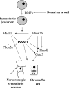
INSM1 in SA lineage transcriptional network. SA precursors are generated from NC cells that give rise to mature neurons of secondary sympathetic ganglia, to chromaffin cells of adrenal medullas, and to extra-adrenal chromaffin tissue. In dorsal aorta wall, the bone morphogenetic proteins BMP2, BMP4, and BMP7 induce Mash1 and Phox2b expression in sympathetic precursors. Genetic studies revealed that Mash1 is upstream of Phox2a and Hand2, whereas Phox2b is upstream of Hand2 and Gata3. Both loss-of-function and gain-of-function experiments indicated reciprocal effects among these factors that form a complex transcriptional network that coordinately regulates differentiation of the SA lineage . An INSM1 mutant mouse study concluded that INSM1 mice showed a reduced proliferation of SA precursor cells that accounted for reduced size of the primary sympathetic ganglion chain and severely impaired terminal differentiation of chromaffin cells .
Figure 5.
Figure 5.
INSM1 displays both cell cycle-dependent and -independent functions. A) INSM1 interacts with cyclin D1 through the cyclin box binding region, which disrupts CDK4 binding and subsequently inhibits Rb protein phosphorylation, E2F release, gene activation, and cell cycle progression. Basically, INSM1 is capable of inducing cell cycle arrest and exit from cell cycle. B) INSM1 exhibits transcriptional repressor activity by binding to the target gene promoter, recruiting cyclin D1 and HDAC-3, and modulating the acetylation status of NeuroD/β2, insulin, and INSM1, the three verified target genes regulated by INSM1.

References

    1. Goto Y, DeSilva M G, Toscani A, Prabhakar B S, Notkins A L, Lan M S. A novel human insulinoma-associated cDNA, IA-1, encodes a protein with zinc-finger DNA-binding motifs. J Biol Chem. 1992;267:15252–15257. - PubMed
    1. Leslie R D G, Atkinson M A, Notkins A L. Autoantigens IA-2 and GAD in type 1 (insulin-dependent) diabetes. Diabetologia. 1999;42:3–14. - PubMed
    1. Lan M S, Li Q, Lu J, Modi W S, Notkins A L. Genomic organization, 5′-upstream sequence, and chromosomal localization of an insulinoma-associated intronless gene, IA-1. J Biol Chem. 1994;269:14170–14174. - PubMed
    1. Shaw G, Kamen R. A conserved AU sequence from the 3′ untranslated region of GM-CSF mRNA mediates selective mRNA degradation. Cell. 1986;46:659–667. - PubMed
    1. Caput D, Beutler B, Hartog K, Thayer R, Brown-Shimer S, Cerami A. Identification of a common nucleotide sequence in the 3′-untranslated region of mRNA molecules specifying inflammatory mediators. Proc Natl Acad Sci U S A. 1986;83:1670–1674. - PMC - PubMed

Publication types

MeSH terms

LinkOut - more resources