. 2009 May 15;18(10):1779-94.
doi: 10.1093/hmg/ddp090. Epub 2009 Feb 26.

Gamma-synucleinopathy: neurodegeneration associated with overexpression of the mouse protein

Affiliations

Gamma-synucleinopathy: neurodegeneration associated with overexpression of the mouse protein

Natalia Ninkina et al. Hum Mol Genet. .

Abstract

The role of alpha-synuclein in pathogenesis of familial and idiopathic forms of Parkinson's disease, and other human disorders known as alpha-synucleinopathies, is well established. In contrast, the involvement of two other members of the synuclein family, beta-synuclein and gamma-synuclein, in the development and progression of neurodegeneration is poorly studied. However, there is a growing body of evidence that alpha-synuclein and beta-synuclein have opposite neuropathophysiological effects. Unlike alpha-synuclein, overexpressed beta-synuclein does not cause pathological changes in the nervous system of transgenic mice and even ameliorates the pathology caused by overexpressed alpha-synuclein. To assess the consequences of excess expression of the third family member, gamma-synuclein, on the nervous system we generated transgenic mice expressing high levels of mouse gamma-synuclein under control of Thy-1 promoter. These animals develop severe age- and transgene dose-dependent neuropathology, motor deficits and die prematurely. Histopathological changes include aggregation of gamma-synuclein, accumulation of various inclusions in neuronal cell bodies and processes, and astrogliosis. These changes are seen throughout the nervous system but are most prominent in the spinal cord where they lead to loss of spinal motor neurons. Our data suggest that down-regulation of small heat shock protein HSPB1 and disintegration of neurofilament network play a role in motor neurons dysfunction and death. These findings demonstrate that gamma-synuclein can be involved in neuropathophysiological changes and the death of susceptible neurons suggesting the necessity of further investigations of the potential role of this synuclein in disease.


Figures

Figure 1.
Figure 1.
Expression of γ-synuclein in wild-type and transgenic mice. (A) A map of DNA fragment used for producing transgenic animals. A mouse γ-synuclein cDNA fragment was inserted between exons II and IV of the mouse Thy-1 gene. (B) Expression of γ-synuclein mRNA in neuronal tissues of wild-type and transgenic animals measured by quantitative real-time RT–PCR. Bar charts show the fold γ-synuclein mRNA level increase (mean ± SEM) in the spinal cord of homozygous and heterozygous 8-week-old Thy1mγSN mice compared with the level in the trigeminal ganglion of newborn wild-type mice (top chart) and in the DRG and spinal cord of homozygous 12-month-old Thy1mγSN mice compared with the level in the DRG of 12-month-old wild-type mice (bottom chart). (C) Western blot analysis of γ-synuclein in the spinal cord of 1-year-old wild-type and homozygous Thy1mγSN animals. The top panel shows a normalization western blot simultaneously probed with antibodies against GAPDH and γ-synuclein. The same undiluted wild-type samples and 1:1000, 1:500 or 1:250 diluted transgenic samples were analysed on a western blot shown in the bottom panel. (D) Western blot analysis of three synucleins in the spinal cord and brain regions of 1-year-old wild-type and Thy1mγSN transgenic (hemi- and homozygous) animals. More γ-synuclein accumulates in neuronal tissues of homozygous than hemizygous mice but a ladder of multimeric γ-synuclein-positive bands is not observed even on overexposed western blots (top panels).
Figure 2.
Figure 2.
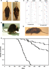
Transgenic animals develop pathology that leads to premature death. Typical appearance of Thy1mγSN transgenic mice at the initial stage of pathology development with clasping of all limbs when hanging by the tail (A) and a hunchback posture (B). Examples of severe gait abnormality in 9- and 12-month-old homozygous γ-synuclein transgenic mice detected by the footprint test. Forefeet prints are blue and hindfeet prints are red (C). At the advanced stage of pathology, the righting reflex is substantially impaired (D). Kaplan–Meier plot of wild-type (squares, n = 49), hemizygous Thy1mγSN (triangles, n = 87) and homozygous Thy1mγSN (circles, n = 74) mice survival over the 26 months period (E).
Figure 3.
Figure 3.
Rotarod performance of γ-synuclein transgenic mice. Groups of mice of different ages and genotypes were tested on Ugo Basile rotarod using constant speed (A) or accelerating (B) modes as described in Material and methods. Line graphs show mean ± SEM of latency to fall for wild-type (diamonds), hemizygous Thy1mγSN (squares) and homozygous (triangles) Thy1mγSN mice at different age. Statistically significant difference (P < 0.01, Student’s t-test) in performance between transgenic and wild type groups at particular age is denoted by asterisks.
Figure 4.
Figure 4.
γ-Synuclein-positive aggregates and fibrils in the spinal cord of 12-month-old transgenic mice detected by anti-mouse γ-synuclein SK23 antibody. (A) Immunostained transverse sections through the spinal cord of wild-type and homozygous γ-synuclein transgenic mice. Higher magnification images of the ventral horn region are shown on lower panels, scale bars = 50 µm. A γ-synuclein-positive cytoplasmic inclusion (further magnified in the left inset) is indicated by the small arrowheads, spheroids by large arrowheads, dystrophic and ballooned neurites by small and large arrows, correspondingly. The right insert shows a hematoxylin and eosin-stained spinal cord motor neuron with an eosinophilic cytoplasmic inclusion, scale bar = 20 µm. (B) Western blot analysis of γ-synuclein in high salt (HS), HS/Triton X-100 (HS/T), RIPA- and SDS-soluble fractions of the spinal cord of wild-type, hemizygous and homozygous mice. (C) Immuno-electron microscopy detection of γ-synuclein fibrils in a sarcosyl-insoluble fraction of the homozygous Thy1mγSN mouse spinal cord.
Figure 5.
Figure 5.
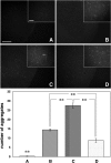
Amyloid deposits in the spinal cord of γ-synuclein transgenic mice. Representative images of Congo Red-stained spinal cord sections of 12-month-old wild-type mouse (A), hemizygous Thy1mγSN mouse (B), homozygous Thy1mγSN mouse at the advance stage of pathology (C) and homozygous Thy1mγSN mouse with mild signs of pathology (D) are shown. Scale bar = 200 µm for all main panels and 25 µm for all insets. The bar chart shows mean ± SEM. of the number of deposits per randomly selected 0.1 mm2 area of the spinal cord gray matter. Sections for counting [104 for (A), 121 for (B), 93 for (C) and 72 for (D)] were randomly selected throughout the length of cervical, thoracic and lumbar parts of the spinal cord of at least four animals per group. One-way ANOVA with post hoc Fisher’s protected t-test demonstrated significant (**P < 0.01) difference between all four experimental groups.
Figure 6.
Figure 6.
Astrogliosis and ubiquitin-positive inclusions in the spinal cord of γ-synuclein transgenic mice. (A) Transverse sections through the spinal cord of wild-type and severely affected homozygous Thy1mγSN mice immunostained for GFAP. Higher magnification images are shown on lower panels, scale bars = 20 µm. (B) Western blot of total protein extracts from 12-month-old wild-type, hemizygous and homozygous transgenic mice probed with antibodies against γ-synuclein, GFAP and GAPDH as a loading control. (C) Transverse sections through the spinal cord of severely affected homozygous Thy1mγSN mouse immunostained for ubiquitin. Boxed areas of gray and white matter are shown at higher magnification on the right top and right bottom panels, correspondingly. (D) Double immunofluorescent staining of severely affected Thy1mγSN mouse spinal cord section with antibodies against ubiquitin (green) and γ-synuclein (red). Examples of dystrophic neuritis and spheroids positive only for γ-synuclein are marked with large arrows, positive only for ubiquitin—with small arrows and positive for both proteins—with arrowheads. Scale bar = 50 µm.
Figure 7.
Figure 7.
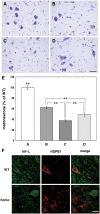
Loss of motor neurons and changes of HSPB1 expression in the spinal cord of γ-synuclein transgenic mice. Representative images of cresyl fast violet-stained spinal cord sections of 12-month-old wild-type mouse (A), hemizygous Thy1mγSN mouse (B), homozygous Thy1mγSN mouse at the advance stage of pathology (C) and homozygous Thy1mγSN mouse with mild pathology (D) are shown. (E) The bar chart shows mean ± SEM. of the number of motor neurons per section expressed as percent of the average number of motor neurons per section of the wild-type mouse spinal cord. Sections for counting [160 for (A), 332 for (B), 215 for (C) and 139 for (D)] were randomly selected throughout the length of cervical, thoracic and lumbar parts of the spinal cord of at least four animals per group. One-way ANOVA with post hoc Fisher’s protected t-test demonstrated significant (**P<0.01) difference between all four experimental groups. (F) Double immunofluorescent staining of spinal cord sections from a wild-type mouse and severely affected homozygous Thy1mγSN mouse with antibodies against neurofilament-L (green) and HSPB1 (red). Scale bar = 20 µm.
Figure 8.
Figure 8.
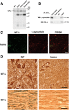
Neurofilaments in the spinal cord and peripheral nerve of γ-synuclein transgenic animals. (A) Western blot analysis of soluble neurofilament-L in the cytosolic (10 000g supernatant) fraction of the spinal cord of 12-month-old wild-type mice, hemizygous Thy1mγSN mice, homozygous Thy1mγSN mice with mild clinical signs of pathology and homozygous Thy1mγSN mice at the advance stage of pathology. (B) Co-immunoprecipitation of γ-synuclein and soluble neurofilament-L from the same cytosolic fractions. (C) Double immunofluorescent staining of severely affected Thy1mγSN mouse spinal cord section with antibodies against neurofilament-L (green) and γ-synuclein (red) shows the absence of NF-L accumulation in γ-synuclein-positive pathological profiles. (D) Substantial reduction of neurofilament-L staining in the ventral horns of the spinal cord (upper panels) and sciatic nerve (lower panels) of 12-month-old homozygous Thy1mγSN mice at the advance stage of pathology compared with their wild-type littermates. Scale bars = 20 µm for (C) and 50 µm for (D).

Similar articles

Cited by

References

    1. Cookson M.R., van der Brug M. Cell systems and the toxic mechanism(s) of alpha-synuclein. Exp. Neurol. 2008;209:5–11. - PMC - PubMed
    1. Park J.Y., Lansbury P.T., Jr Beta-synuclein inhibits formation of alpha-synuclein protofibrils: a possible therapeutic strategy against Parkinson’s disease. Biochemistry. 2003;42:3696–3700. - PubMed
    1. da Costa C.A., Masliah E., Checler F. Beta-synuclein displays an antiapoptotic p53-dependent phenotype and protects neurons from 6-hydroxydopamine-induced caspase 3 activation: cross-talk with alpha-synuclein and implication for Parkinson’s disease. J. Biol. Chem. 2003;278:37330–37335. - PubMed
    1. Windisch M., Hutter-Paier B., Rockenstein E., Hashimoto M., Mallory M., Masliah E. Development of a new treatment for Alzheimer’s disease and Parkinson’s disease using anti-aggregatory beta-synuclein-derived peptides. J. Mol. Neurosci. 2002;19:63–69. - PubMed
    1. Hashimoto M., Rockenstein E., Mante M., Mallory M., Masliah E. β-Synuclein inhibits alpha-synuclein aggregation: a possible role as an anti-parkinsonian factor. Neuron. 2001;32:213–223. - PubMed

Publication types

MeSH terms

Substances