. 2009 Apr;37(7):2346-58.
doi: 10.1093/nar/gkp098. Epub 2009 Feb 26.

Posttranscriptional regulation of angiotensin II type 1 receptor expression by glyceraldehyde 3-phosphate dehydrogenase

Affiliations

Posttranscriptional regulation of angiotensin II type 1 receptor expression by glyceraldehyde 3-phosphate dehydrogenase

Michael Backlund et al. Nucleic Acids Res. 2009 Apr.

Abstract

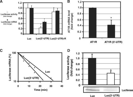
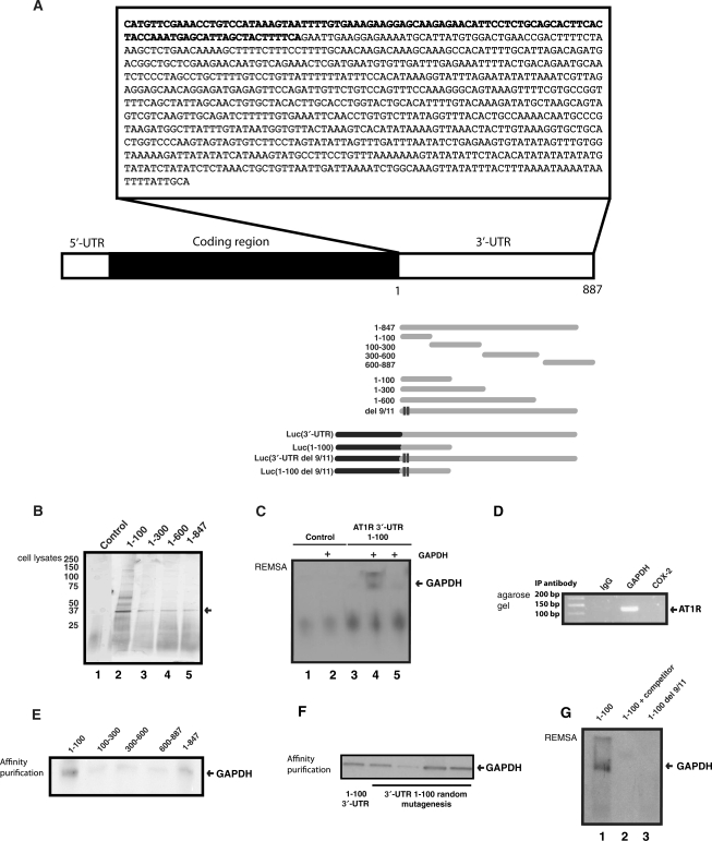
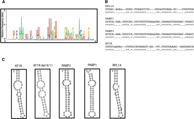
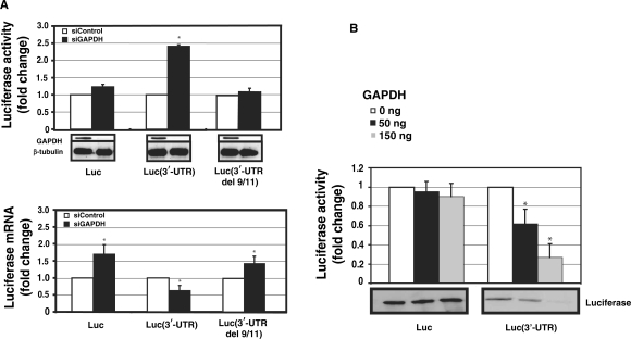
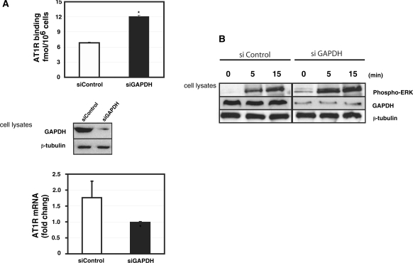
Regulation of angiotensin II type 1 receptor (AT1R) has a pathophysiological role in hypertension, atherosclerosis and heart failure. We started from an observation that the 3'-untranslated region (3'-UTR) of AT1R mRNA suppressed AT1R translation. Using affinity purification for the separation of 3'-UTR-binding proteins and mass spectrometry for their identification, we describe glyceraldehyde 3-phosphate dehydrogenase (GAPDH) as an AT1R 3'-UTR-binding protein. RNA electrophoretic mobility shift analysis with purified GAPDH further demonstrated a direct interaction with the 3'-UTR while GAPDH immunoprecipitation confirmed this interaction with endogenous AT1R mRNA. GAPDH-binding site was mapped to 1-100 of 3'-UTR. GAPDH-bound target mRNAs were identified by expression array hybridization. Analysis of secondary structures shared among GAPDH targets led to the identification of a RNA motif rich in adenines and uracils. Silencing of GAPDH increased the expression of both endogenous and transfected AT1R. Similarly, a decrease in GAPDH expression by H(2)O(2) led to an increased level of AT1R expression. Consistent with GAPDH having a central role in H(2)O(2)-mediated AT1R regulation, both the deletion of GAPDH-binding site and GAPDH overexpression attenuated the effect of H(2)O(2) on AT1R mRNA. Taken together, GAPDH is a translational suppressor of AT1R and mediates the effect of H(2)O(2) on AT1R mRNA.


Figures

Figure 1.
Figure 1.
3′-UTR of AT1R is a negative regulator of AT1R expression. (A) The effect of AT1R 3′-UTR on luciferase activity (white bars). Three different constructs were studied: luciferase, luciferase with AT1R 3′-UTR and luciferase fused with AT1R 3′-UTR in reverse orientation. HEK293 cells were transfected with a luciferase and renilla luciferase plasmids. Cells were lysed and assayed for firefly and renilla luciferase activities. To evaluate mRNA levels, luciferase and GAPDH mRNA levels were measured by quantitative PCR. Corrected luciferase mRNA expression values were calculated by dividing luciferase mRNA by GAPDH mRNA expression (black bars). Values were normalized to the activity or mRNA levels of a luciferase reporter lacking the AT1R 3′-UTR to account for the effect of 3′-UTR. *P < 0.05 versus luciferase without 3′-UTR. (B) The effect of 3′-UTR on AT1R mRNA was evaluated. We compared the mRNA levels of AT1R coding region with or without 3'-UTR. AT1R and GAPDH mRNA levels were determined by quantitative PCR using AT1R coding region specific oligos. AT1R expression was corrected by GAPDH expression. Data were normalized to the activity of AT1R construct lacking the 3′-UTR. The results represent the means ± SD of an average of three independent experiments performed in triplicate for each construct. *P < 0.05 versus AT1R without 3′-UTR. (C) Measurement of the rate of degradation of luciferase mRNA. HEK293 cells were transiently transfected with luciferase construct with or without the full length AT1R 3′-UTR. Transfected cells were then treated with actinomycin D for 5 min, 10 min, 15 min and 30 min. Quantitative PCR was performed with luciferase and GAPDH specific oligos and luciferase mRNA expression was normalized by GAPDH. The mRNA measurements for each construct were normalized to the expression of the construct at time zero. Results are shown as a linear fit. Results represent the means of an average of three independent experiments performed as a triplicate for each construct. (D). Measurement of the rate of luciferase translation in rabbit reticulocyte lysates. In vitro translation of chimeric luciferase constructs with and without 3′-UTR were compared. From the in vitro translation reaction mixture luciferase activity was measured (upper panel) and western blot analysis was performed (lower panel). Translated proteins were detected with streptavidin-HRP detection system. *P < 0.05 versus luciferase without 3′-UTR.
Figure 2.
Figure 2.
GAPDH interacts with AT1R 3′-UTR. (A) A schematic of AT1R mRNA transcript as well as deleted and truncated transcripts used in affinity purification and gel shift assays. (B) Search for RNA-binding proteins that form a complex with AT1R 3′-UTR. The details of the purification are given under ‘Materials and Methods’ section. Lane 1: Luciferase coding region template was used as a control. Lanes 2–5: In vitro transcribed truncated fragments of AT1R 3'-UTR were used in the affinity purification. The differentially expressed 36 kDa protein was excised, digested, and the resulting peptides were recognized by mass spectrometry. (C) REMSA. After binding reaction, excess unbound RNA was digested. In lane 1A biotinylated control probe of luciferase coding region without GAPDH was used. Lane 2 has the same control probe as lane 1 but 150 ng of purified GAPDH protein was added. Lane 3 had biotinylated 3′-UTR 1–100 transcript without GAPDH, lane 4 had 150 ng of GAPDH and lane 5 had both 150 ng of GAPDH as well as 100-fold excess of unlabelled probe included in the binding mixture. (D) Binding of endogenous GAPDH with endogenous AT1R mRNA was detected by RT-qPCR assay with AT1R specific primers of material obtained by IP from cytoplasmic fractions. Changes in the level of AT1R mRNA associated with GAPDH were evaluated by measuring its abundance in the IP mixture. Immunoprecipitation was performed with preimmuno IgG, with GAPDH-specific antibody, and with anti-cox-2 antibody. PCR products were visualized after electrophoresis in 1% agarose gels stained with ethidium bromide. (E) Mapping of the GAPDH-binding site within AT1R 3′-UTR. A western blot of proteins isolated by RNA affinity purification was performed and GAPDH expression was detected by polyclonal anti-GAPDH antibodies. (F) Random mutagenesis of the 1–100 part of AT1R 3′-UTR. Mutated 3′-UTRs were generated by PCR-based mutagenesis. One hundred clones were generated and of those ten were sequenced to determine the rate of mutagenesis. Each mutant had 2 to 4 base insertions, deletions or mutations. We measured the binding of the endogenous GAPDH to the mutants by affinity purification. To reduce variability, the same HEK 293 cytoplasmic cell lysate was used for all these constructs. After protein binding, RNA treatment was performed. (G) REMSA of AT1R 3′-UTR Del 9/11. Lane 1. A biotinylated probe of 1–100 was used as a positive control for GAPDH REMSA. Cell lysate was from HEK293 cells. Lane 2. A biotinylated probe of 1–100 was used with 10-fold excess of unlabelled probe included in the binding mixture. Lane 3. A biotinylated transcript containing 1–100 del 9/11 that lacks GAPDH-binding site.
Figure 3.
Figure 3.
Sequence and structure of the predicted GAPDH motif, as identified among GAPDH-bound transcripts. (A) Probability matrix (graphic logo) of the GAPDH motif indicating the relative frequency of finding each residue at each position within the motif, as elucidated from the array-derived experimental data set. (B) Structural alignment of four examples of GAPDH motif in specific mRNAs. (C) Secondary structure of four representative examples of the GAPDH motif in specific mRNAs. RPL14, ribosomal protein L14; PABP1, poly (A)-binding protein 1; PABP3, poly (A)-binding protein 3; AT1R, angiotensin II type 1 receptor. Furthermore, del 9/11 of AT1R 3′-UTR is shown.
Figure 4.
Figure 4.
The effect of RNAi-mediated GAPDH knockdown on the expression of AT1R. HEK293 cells were cotransfected with luciferase vector and with 30 nM gene-specific GAPDH siRNA (black bars) or equal amount of control siRNA (white bars). (A) Upper panel: Luciferase activities of luciferase coding region only or fused with full length AT1R 3′-UTR or mutated full-length 3′-UTR del 9/11. Luminometer measurements for this and subsequent experiments are expressed as fold of promoter control without 3′-UTR in relative light units of firefly luciferase. Results represent the means ± SD of an average of three independent experiments performed as a triplicate for each construct. β-Tubulin was measured to control for protein loading. *P < 0.05 versus siControl-treated cells. Lower panel: Luciferase mRNA was quantified by quantitative PCR. All the results were normalized to β-actin expression. *P < 0.05 versus siControl transfected cells. (B) GAPDH suppresses AT1R 3′-UTR translation. The same transcripts as in Figure 1 were used. Increasing amounts of GAPDH was added to the reaction mixture (white bars 0 ng, black bars 50 ng and gray bars 150 ng). The translated product was evaluated by two methods. First, luciferase activity of the translation mixture was measured (upper panel). Second, lysate was separated on SDS–PAGE gel, blotted and detected on a membrane by streptavidin-HRP (lower panel). Experiment with and without AT1R 3′-UTR represent different exposures and therefore baseline difference between these two constructs cannot be compared. The results represent the means ± SD of an average of three independent experiments. *P < 0.05 versus control reaction with no GAPDH.
Figure 5.
Figure 5.
Role of GAPDH in the regulation of endogenous AT1R. (A) Upper panel: The effect of GAPDH silencing on AT1R expression in early passage (3–5) coronary artery VSMCs. VSMCs were transfected with 30 nM GAPDH siRNA or equal amount of control siRNA. AT1R expression was quantified by ligand binding. The western blot below demonstrates the effect of GAPDH siRNA on GAPDH expression. Lower panel: AT1R mRNA was quantified by RT–qPCR. All the results were normalized to β-actin expression. *P < 0.05 versus siControl transfected cells. (B) Activation of ERK MAPKs in human coronary artery VSMC by angiotensin II. Coronary artery VSMC were transfected either with siGAPDH or control siRNA. These cells were serum starved for 24 h before stimulation by 10−7 M angiotensin II. Cells were flash frozen at appropriate time points, proteins extracted and results analyzed by western blotting using anti-phospho-ERK antibody, GAPDH antibodies, and β-tubulin antibody.
Figure 6.
Figure 6.
(A) Effect of H2O2 on AT1R expression in human coronary artery VSMC. These VSMC were exposed to varying concentrations of H2O2 for 15 h. Western blots were probed with appropriate antibodies. (B) GAPDH binding to AT1R 3′-UTR was evaluated by affinity purification using the 3′-UTR 0–847 as a probe (same as in Figure 2B and E) and western blotting of the isolated proteins by GAPDH antibodies. GAPDH expression was evaluated in total cellular lysates prepared from HEK293 cells exposed to 150 μM H2O2 for 15 h (lower panel, right). β-Tubulin was measured to control for protein loading. (C) H2O2 treatment reduces GAPDH binding to AT1R. Exposure of protein lysates to H2O2 does not influence protein levels but decreases GAPDH binding to 3′-UTR in a dose-dependent manner. Left side of the REMSA has a control blot (lanes 1–2) and the right side has the experiment (lanes 3–5) in which protein lysate was exposed to H2O2 for 1 h. (D) GAPDH mediates H2O2-induced effect on the posttranscriptional regulation of AT1R. The same luciferase constructs as in Figure 4A were used. HEK293 cells were transfected with either β-galactosidase (left panel) or GAPDH expression vector (right panel) together with luciferase constructs with (3′-UTR, 1–100) or without GAPDH-binding site (no 3′-UTR, 1–100 del 9/11). Cells were exposed to 150 μM H2O2 for 15 h (black bar) or to vehicle (white bar), lysed and assayed for luciferase activities. Values were normalized to the activity of untreated luciferase construct. The results represent the means ± SD of an average of three independent experiments performed in triplicate for each construct. *P < 0.05 versus AT1R without 3′-UTR.

Similar articles

Cited by

References

    1. Dzau V. The cardiovascular continuum and renin-angiotensin-aldosterone system blockade. J. Hypertens. Suppl. 2005;23:S9–S17. - PubMed
    1. Horiuchi M, Akishita M, Dzau VJ. Recent progress in angiotensin II type 2 receptor research in the cardiovascular system. Hypertension. 1999;33:613–621. - PubMed
    1. Lehtonen JY, Daviet L, Nahmias C, Horiuchi M, Dzau VJ. Analysis of functional domains of angiotensin II type 2 receptor involved in apoptosis. Mol. Endocrinol. 1999;13:1051–1060. - PubMed
    1. Daviet L, Lehtonen JY, Tamura K, Griese DP, Horiuchi M, Dzau VJ. Cloning and characterization of ATRAP, a novel protein that interacts with the angiotensin II type 1 receptor. J. Biol. Chem. 1999;274:17058–17062. - PubMed
    1. Elton TS, Martin MM. Angiotensin II type 1 receptor gene regulation: transcriptional and posttranscriptional mechanisms. Hypertension. 2007;49:953–961. - PubMed

Publication types

MeSH terms