. 2009 May 14;113(20):4914-7.
doi: 10.1182/blood-2008-11-189845. Epub 2009 Feb 26.

Deregulation of microRNA involved in hematopoiesis and the immune response in HTLV-I adult T-cell leukemia

Affiliations

Deregulation of microRNA involved in hematopoiesis and the immune response in HTLV-I adult T-cell leukemia

Marcia Bellon et al. Blood. .

Abstract

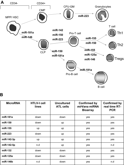
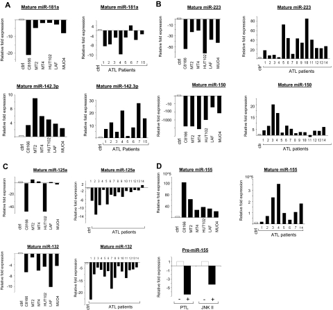
Human T-cell leukemia virus type-I (HTLV-I) is the etiologic agent of adult T-cell leukemia (ATL), an aggressive lymphoproliferative disease. MicroRNAs (miRNAs) are differentially expressed during hematopoiesis and lineage commitment of hematopoietic stem cell progenitors (HSCPs). Here, we report aberrant expression of hematopoietic-specific miR-223, miR-181a, miR-150, miR-142.3p, and miR-155 in HTLV-I-infected cells in vitro and uncultured ex vivo ATL cells. Our results suggest that HTLV-I-infected cells have an unbalanced expression of miRNA that favors T-cell differentiation. We also found altered expression of miRNA previously recognized as innate immunity regulators: miR-155, miR-125a, miR-132, and miR-146. Strikingly, our data also revealed significant differences between ex vivo ATL tumor cells and in vitro HTLV-I cell lines. Specifically, miR-150 and miR-223 were up-regulated in ATL patients but consistently down-regulated in HTLV-I cell lines, suggesting that ATL cells and in vitro-established cells are derived from distinct cellular populations.


Figures

Figure 1
Figure 1
Deregulation of miRNA involved in hematopoiesis and innate immunity in HTLV-I–infected cells. (A) miRNA involved in the different steps of hematopoiesis and showing deregulated expression in HTLV-I–transformed cells in vitro and ATL tumor cells ex vivo. MPP indicates myeloid pluripotent progenitors, HSC, hematopoietic stem cells; CMP, common myeloid progenitors; CLP, common lymphocyte progenitors; and CFU-GM, colony-forming units granulocyte-macrophage. (B) Table representation of mature miRNA deregulated in HTLV-I–infected cells in vitro and ex vivo identified by mirVana miRNA Bioarrays platform and real-time RT-PCR. The miRNA enriched fraction was obtained by passing total RNA through a flashPAGE Fractionator apparatus (Ambion, Austin, TX). The 3′ ends of the RNA molecules were tailed and labeled using the mirVana miRNA Labeling Kit (Ambion) and analyzed as described in “Methods.” nd indicates not determined.
Figure 2
Figure 2
Real-time quantification of mature miRNA in HTLV-I cell lines and ex vivo ATL samples. (A-D) TaqMan real-time RT-PCR from HTLV-I–transformed cells (C8166, MT2, MT4, and HUT102) and immortalized cells (LAF and MU04) in vitro and ATL tumor cells ex vivo. The TaqMan probes that were used ensure accurate discrimination between miRNAs that may differ by a single nucleotide. Mature miRNAs were analyzed because maturation of pri-miRNA is subject to posttranscriptional regulations. The threshold cycle (CT) is defined as the fractional cycle number at which the fluorescence passes the fixed threshold. TaqMan CT values are used to measure the fold change. ATL samples and HTLV-I cell lines were compared with peripheral blood mononuclear cells (PBMCs) and purified CD4 T cells as controls. miR-24 was used as the internal control, because of its constitutive expression among ATL samples, control T cells, and HTLV-I cell lines. These experiments were performed by Asuragen (Austin, TX). All primers and probes were tested and validated by Asuragen. Statistical analysis is provided in Document S1. For panel D, MT4 cells were treated overnight with either 6 μM parthenolide (PTL) or 90 μM JNK II. Control cells were treated with DMSO. SYBR green real-time PCR was performed for pre-miR-155 expression (F: 5′-CTGTTAATGCTAATCGTGATAG-3′ and R: 5′-AATGCTAATATGTAGGAGTCAG-3′) with GAPDH (F: 5′-GAAGGTGAAGGTCGGAGTC-3′ and R: 5′-GAAGATGGTGATGGGATTTC-3′) as an internal control. All experiments were performed in triplicate.

References

    1. Calin GA, Croce CM. MicroRNA signatures in human cancers. Nat Rev Cancer. 2006;6:857–866. - PubMed
    1. Lu J, Getz G, Miska EA, et al. MicroRNA expression profiles classify human cancers. Nature. 2005;435:834–838. - PubMed
    1. Kluiver J, Kroesen BJ, Poppema S, van den BA. The role of microRNAs in normal hematopoiesis and hematopoietic malignancies. Leukemia. 2006;20:1931–1936. - PubMed
    1. García P, Frampton J. Hematopoietic lineage commitment: miRNAs add specificity to a widely expressed transcription factor. Dev Cell. 2008;14:815–816. - PubMed
    1. Garzon R, Croce CM. MicroRNAs in normal and malignant hematopoiesis. Curr Opin Hematol. 2008;15:352–358. - PubMed

Publication types

MeSH terms