

One tissue, two fates: different roles of megagametophyte cells during Scots pine embryogenesis
- PMID: 19246593
- PMCID: PMC2657542
- DOI: 10.1093/jxb/erp020
One tissue, two fates: different roles of megagametophyte cells during Scots pine embryogenesis
Abstract
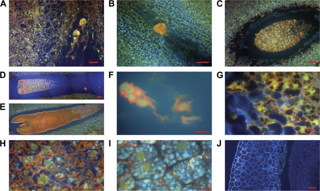
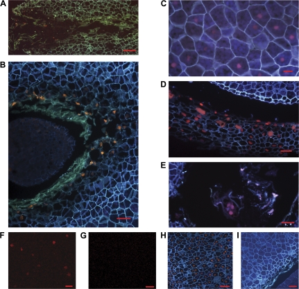
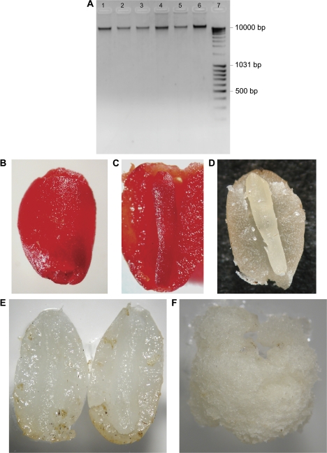
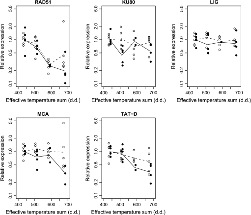
In the Scots pine (Pinus sylvestris L.) seed, embryos grow and develop within the corrosion cavity of the megagametophyte, a maternally derived haploid tissue, which houses the majority of the storage reserves of the seed. In the present study, histochemical methods and quantification of the expression levels of the programmed cell death (PCD) and DNA repair processes related genes (MCA, TAT-D, RAD51, KU80, and LIG) were used to investigate the physiological events occurring in the megagametophyte tissue during embryo development. It was found that the megagametophyte was viable from the early phases of embryo development until the early germination of mature seeds. However, the megagametophyte cells in the narrow embryo surrounding region (ESR) were destroyed by cell death with morphologically necrotic features. Their cell wall, plasma membrane, and nuclear envelope broke down with the release of cell debris and nucleic acids into the corrosion cavity. The occurrence of necrotic-like cell death in gymnosperm embryogenesis provides a favourable model for the study of developmental cell death with necrotic-like morphology and suggests that the mechanism underlying necrotic cell death is evolutionary conserved.
Figures
Similar articles
-
Pine embryogenesis: many licences to kill for a new life.Plant Signal Behav. 2009 Oct;4(10):928-32. doi: 10.4161/psb.4.10.9535. Epub 2009 Oct 16. Plant Signal Behav. 2009. PMID: 19826239 Free PMC article. Review.
-
Microscopical Detection of Cell Death Processes During Scots Pine Zygotic Embryogenesis.Methods Mol Biol. 2020;2122:223-237. doi: 10.1007/978-1-0716-0342-0_16. Methods Mol Biol. 2020. PMID: 31975306
-
Expression of catalase and retinoblastoma-related protein genes associates with cell death processes in Scots pine zygotic embryogenesis.BMC Plant Biol. 2015 Mar 15;15:88. doi: 10.1186/s12870-015-0462-0. BMC Plant Biol. 2015. PMID: 25887788 Free PMC article.
-
Moderate stress responses and specific changes in polyamine metabolism characterize Scots pine somatic embryogenesis.Tree Physiol. 2016 Mar;36(3):392-402. doi: 10.1093/treephys/tpv136. Epub 2016 Jan 19. Tree Physiol. 2016. PMID: 26786537 Free PMC article.
-
Programmed cell death in plant embryogenesis.Curr Top Dev Biol. 2005;67:135-79. doi: 10.1016/S0070-2153(05)67004-4. Curr Top Dev Biol. 2005. PMID: 15949533 Review.
Cited by
-
Detection of De Novo Mutations by Sequencing Reduced Representation Libraries.Methods Mol Biol. 2025;2933:99-111. doi: 10.1007/978-1-0716-4574-1_14. Methods Mol Biol. 2025. PMID: 40418480
-
Cryo-Treatment Enhances the Embryogenicity of Mature Somatic Embryos via the lncRNA-miRNA-mRNA Network in White Spruce.Int J Mol Sci. 2022 Jan 20;23(3):1111. doi: 10.3390/ijms23031111. Int J Mol Sci. 2022. PMID: 35163033 Free PMC article.
-
Pine embryogenesis: many licences to kill for a new life.Plant Signal Behav. 2009 Oct;4(10):928-32. doi: 10.4161/psb.4.10.9535. Epub 2009 Oct 16. Plant Signal Behav. 2009. PMID: 19826239 Free PMC article. Review.
-
Mitochondrial bioenergetics linked to the manifestation of programmed cell death during somatic embryogenesis of Abies alba.Planta. 2009 Dec;231(1):93-107. doi: 10.1007/s00425-009-1028-x. Epub 2009 Oct 16. Planta. 2009. PMID: 19834734
-
Dealing with the problem of non-specific in situ mRNA hybridization signals associated with plant tissues undergoing programmed cell death.Plant Methods. 2010 Feb 5;6(1):7. doi: 10.1186/1746-4811-6-7. Plant Methods. 2010. PMID: 20181098 Free PMC article.
References
-
- Ameisen JC. On the origin, evolution, and nature of programmed cell death: a timeline of four billion years. Cell Death and Differentiation. 2002;9:367–393. - PubMed
-
- Ann W, Syring J, Gernandt DS, Liston A, Cronn R. Fossil calibration of molecular divergence infers a moderate mutation rate and recent radiations for Pinus. Molecular Biology and Evolution. 2007;24:90–101. - PubMed
-
- Barzilai A, Yamamoto KI. DNA damage responses to oxidative stress. DNA Repair. 2004;3:1109–1115. - PubMed
-
- Becwar MR, Nagmani R, Wann SR. Initiation of embryogenic cultures and somatic embryo development in loblolly pine (Pinus taeda) Canadian Journal of Forest Research. 1990;20:810–817.
-
- Beers EP. Programmed cell death during plant growth and development. Cell Death and Differentiation. 1997;4:649–661. - PubMed
Publication types
MeSH terms
Substances
LinkOut - more resources
Full Text Sources
Research Materials
Miscellaneous
